
Radiographic image processing device, method of operating radiographic image processing device, and radiographic image processing program

a radiographic image and processing device technology, applied in image analysis, medical science, diagnostics, etc., can solve problems such as the deterioration of the contrast of radiographic images

Active Publication Date: 2020-10-01
FUJIFILM CORP
View PDF0 Cites 0 Cited by
  • Summary
  • Abstract
  • Description
  • Claims
  • Application Information

AI Technical Summary

Benefits of technology

The invention is a device, method, and program that can estimate the unwanted radiation that reaches a detection unit without passing through a patient. This estimation can then be used to remove the unwanted radiation to improve the image quality of the detected radiation.

Problems solved by technology

The scattered radiation of the direct radiation region also causes the contrast of the radiographic image to deteriorate.

Method used


Image

  • Radiographic image processing device, method of operating radiographic image processing device, and radiographic image processing program
  • Radiographic image processing device, method of operating radiographic image processing device, and radiographic image processing program
  • Radiographic image processing device, method of operating radiographic image processing device, and radiographic image processing program

Examples


Embodiment Construction

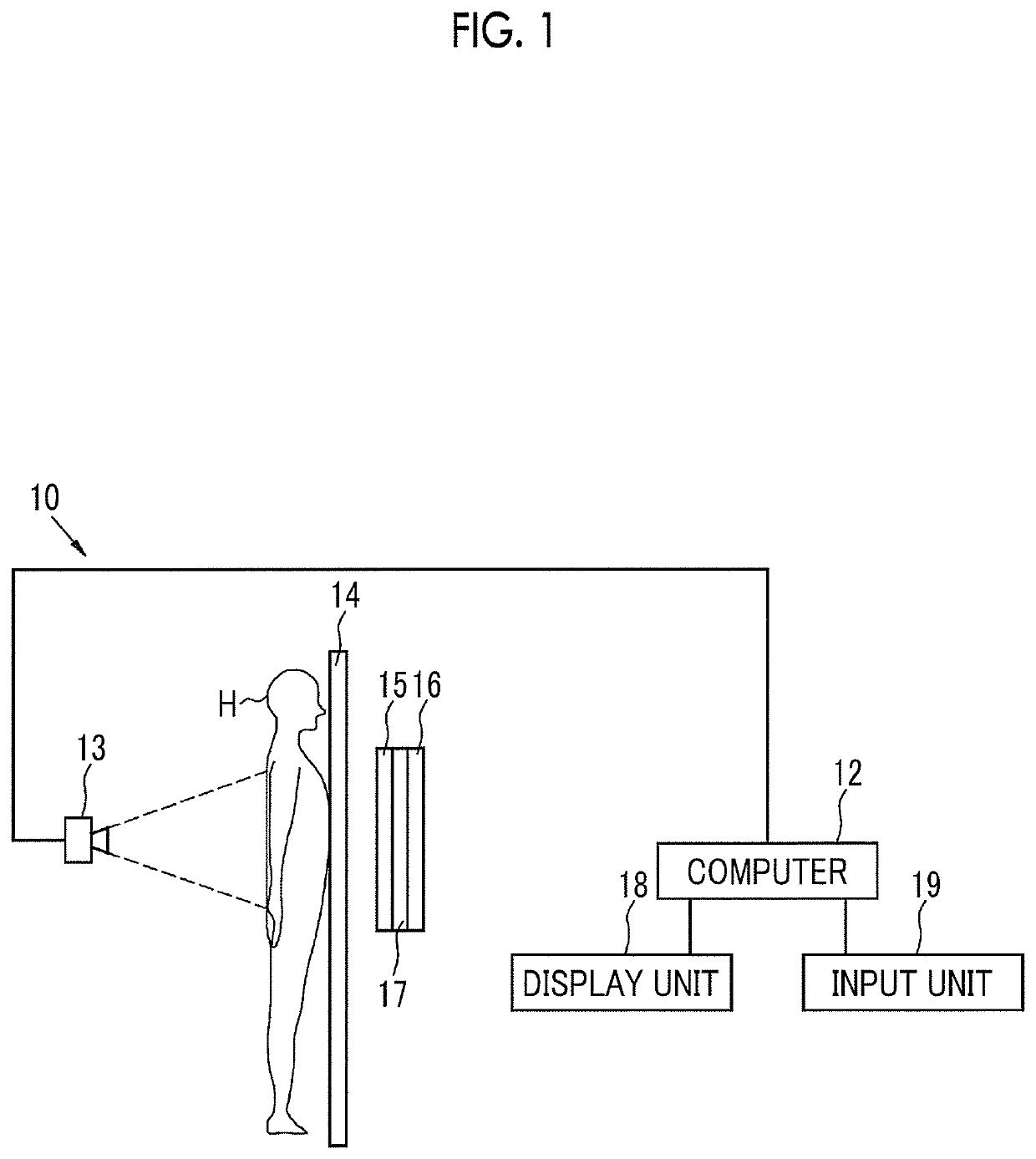
[0025]As shown in FIG. 1, a radiographic imaging system comprises an imaging device 10 and a computer 12 and acquires two radiographic images having different energy distributions. In a case where the imaging device 10 receives radiation (for example, X-ray), which is emitted from a radiation source 13 and is transmitted through a subject H, through a top board 14 by a first radiation detector 15 and a second radiation detector 16, each of the first and second radiation detectors 15 and 16 converts the radiation into energy and receives the energy (one-shot energy subtraction). The top board 14, the first radiation detector 15, a radiation energy conversion filter 17 formed of a copper plate or the like, and the second radiation detector 16 are arranged in this order from the side close to the radiation source 13 and the radiation source 13 is driven at the time of imaging. The first and second radiation detectors 15 and 16 and the radiation energy conversion filter 17 are in close ...


PUM


Abstract

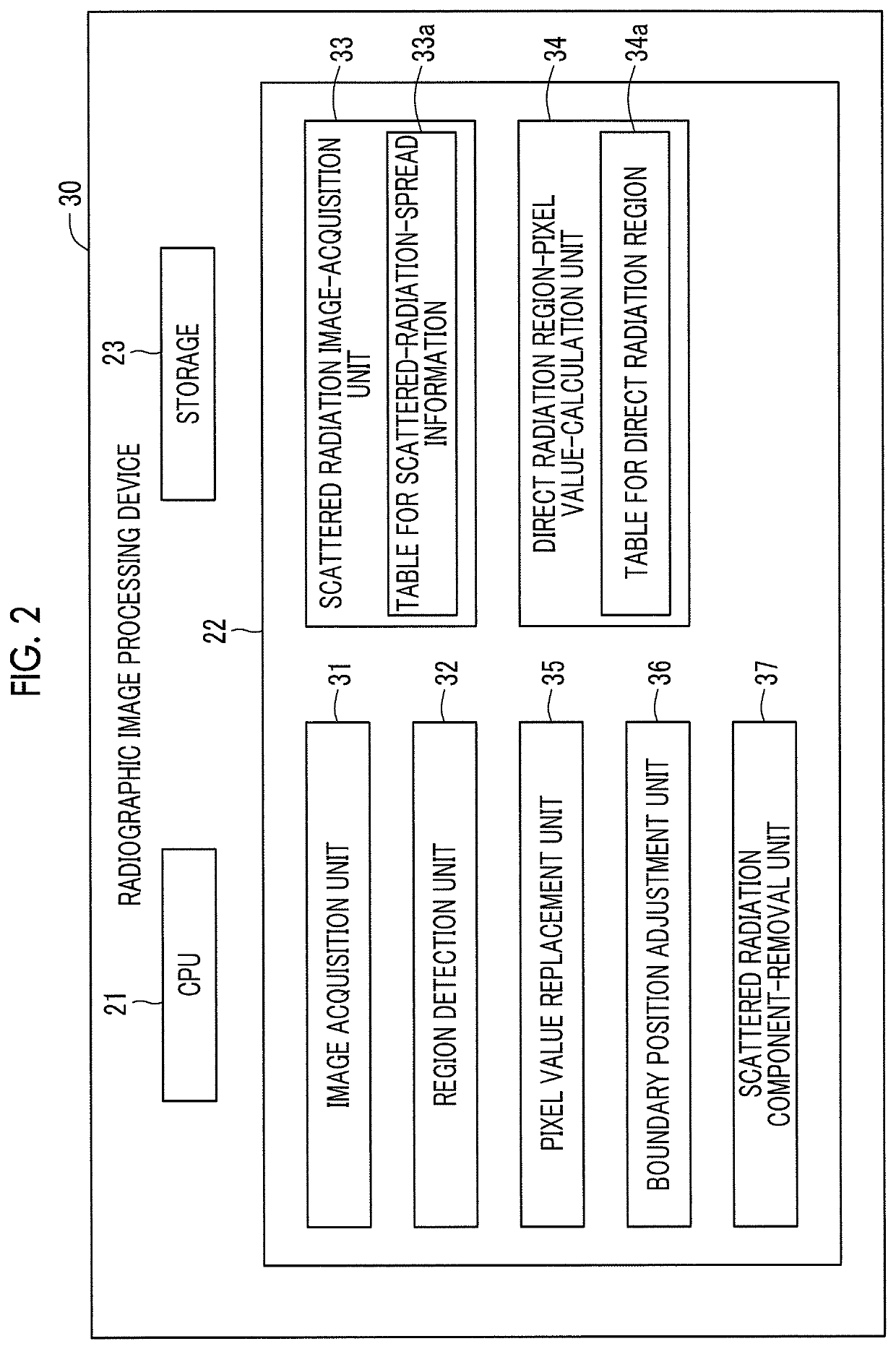
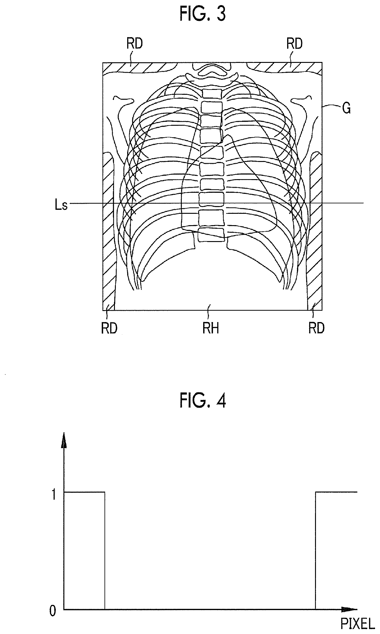
A region detection image is obtained by the detection of a subject region of a radiographic image in which radiation is transmitted through a subject and reaches a radiation detection unit and a direct radiation region of the radiographic image in which the radiation directly reaches the radiation detection unit without being transmitted through the subject. A scattered radiation image about scattered radiation components is obtained on the basis of the region detection image and scattered-radiation-spread information about the spread of scattered radiation. A radiographic image from which the scattered radiation components have been removed is obtained by the subtraction of the scattered radiation image from the radiographic image.

Description

CROSS-REFERENCE TO RELATED APPLICATIONS[0001]This application claims priority under 35 U.S.0§ 119(a) to Japanese Patent Application No. 2019-066182 filed on 29 Mar. 2019. The above application is hereby expressly incorporated by reference, in its entirety, into the present application.BACKGROUND OF THE INVENTION1. Field of the Invention[0002]The present invention relates to a radiographic image processing device, a method of operating the radiographic image processing device, and a radiographic image processing program that process a radiographic image obtained from the imaging of a subject irradiated with radiation.2. Description of the Related Art[0003]A diagnosis, which uses a radiographic image obtained in a case where a subject irradiated with radiation is imaged by a radiation detection unit, is made in a medical field. Scattered radiation causing the contrast of a subject to deteriorate is included in radiation that is transmitted through the subject and reaches the radiation...

Claims


Application Information

Patent Timeline
Patent Type & Authority Applications(United States)
IPC IPC(8): A61B6/00G06T7/00
CPCA61B6/465A61B6/54A61B6/5282G06T7/0012G06T2207/10116G06T5/50A61B6/482A61B6/469A61B6/4291
Inventor TAKAHASHI, TOMOYUKI
Owner FUJIFILM CORP
Learn More