
Device and method for obtaining anatomical measurements from an ultrasound image

an ultrasound image and anatomical measurement technology, applied in image enhancement, instruments, catheters, etc., can solve the problems of high labor intensity, high labor intensity, and inability to meet the preferences of sonographers, so as to save operator time and effort, improve efficiency, and improve robustness

Pending Publication Date: 2021-03-18
KONINKLJIJKE PHILIPS NV
View PDF0 Cites 1 Cited by
  • Summary
  • Abstract
  • Description
  • Claims
  • Application Information

AI Technical Summary

Benefits of technology

The invention is about a method for automatically detecting and measuring in an image based on the location and geometrical characteristics of the set of points selected by an operator. This saves time and effort for the operator by avoiding the need to manually select the measurement type in advance. The method is designed to improve efficiency and flexibility over standard approaches, while retaining the clinician's key expertise and ensuring clinical robustness. The method also provides a way for the operator to decide ad hoc which measurements to do and in which order, and the method is able to response appropriately and apply the appropriate tool.

Problems solved by technology

If multiple views are to be generated with such a procedure, there is a risk of missed abnormalities because obtaining and analyzing these views requires high skill (e.g. fetal echocardiography is very operator-dependent) whilst in addition the fetus may be moving during the procedure, requiring the clinician to reorient himself or herself with the fetus each time the fetus moves.
This workflow is slow and tedious, as it requires the operator to select each measurement to be made from a long list before it can be performed.
However, it is highly restrictive to the sonographer and may not suit their preferences.
It also fails to provide flexibility for performing additional measurements which may be clinically appropriate in given situations.
While these systems reduce workload, they suffer from lack of robustness, and are never able to achieve 100% accuracy.
This also makes achieving regulatory approval for their use in devices very difficult, since their reliability is low.

Method used


Image

  • Device and method for obtaining anatomical measurements from an ultrasound image
  • Device and method for obtaining anatomical measurements from an ultrasound image
  • Device and method for obtaining anatomical measurements from an ultrasound image

Examples


Embodiment Construction

[0072]The invention will be described with reference to the Figures.

[0073]It should be understood that the detailed description and specific examples, while indicating exemplary embodiments of the apparatus, systems and methods, are intended for purposes of illustration only and are not intended to limit the scope of the invention. These and other features, aspects, and advantages of the apparatus, systems and methods of the present invention will become better understood from the following description, appended claims, and accompanying drawings. It should be understood that the Figures are merely schematic and are not drawn to scale. It should also be understood that the same reference numerals are used throughout the Figures to indicate the same or similar parts.

[0074]The invention provides a method for making anatomical measurements using a captured ultrasound image representative of the anatomical region of interest. The method comprises receiving data representative of a set of...


PUM


Abstract

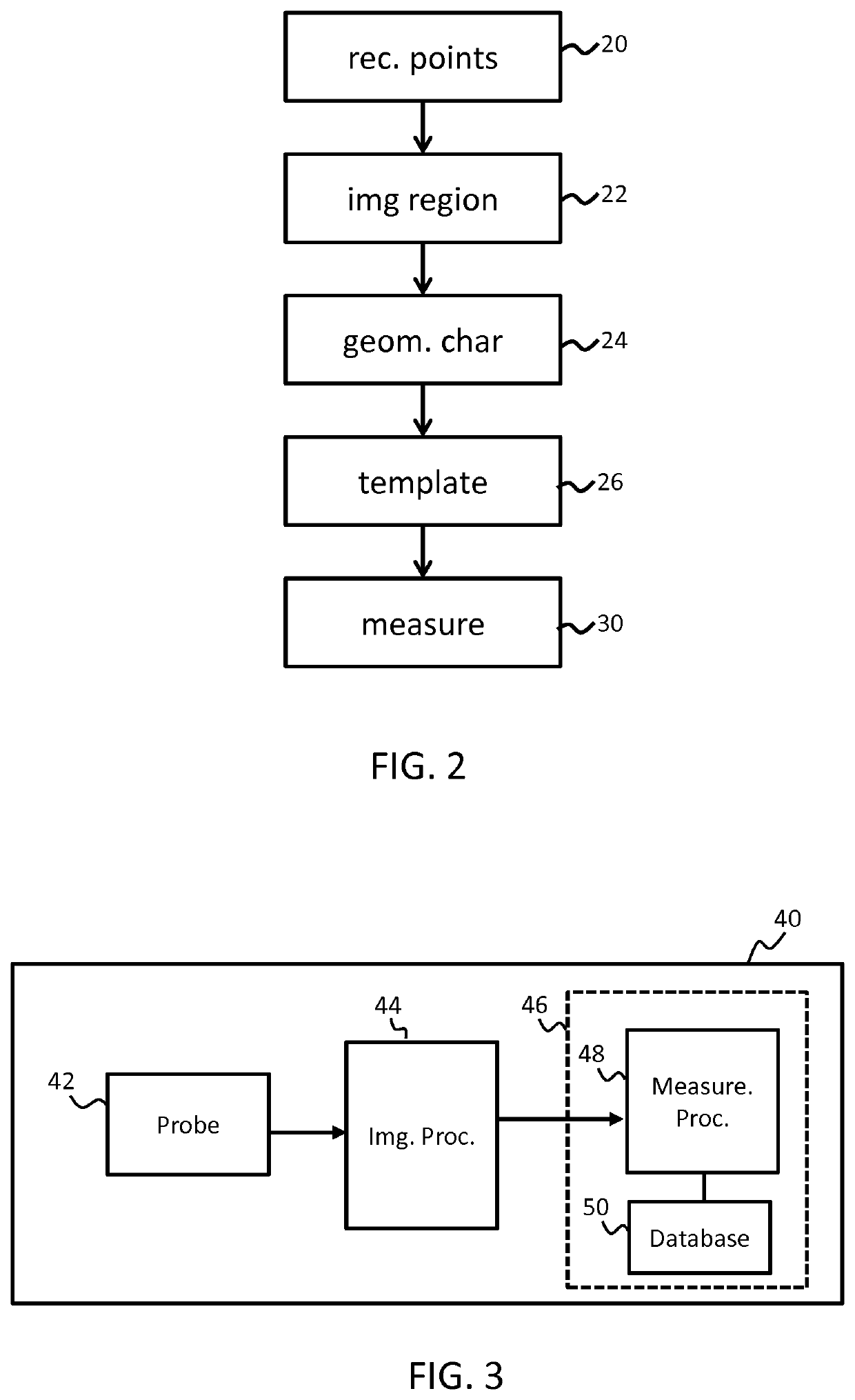
The invention provides a method for making anatomical measurements using a captured ultrasound image representative of the anatomical region of interest. The method comprises receiving (20) data representative of a set of points selected by a user within the image and interpreting from the points a particular anatomical measurement type which the user is intending to perform. In particular, the points are processed to identify (24) a pattern or other geometrical characteristic of the points, and the location (22) of at least one of the points within the image is also identified. These two characteristics are used to identify from the set of points which measurement the operator is performing. Once the measurement is identified, an appropriate measurement template (26) is selected and applied in order to derive (30) from the set of points an anatomical measurement.

Description

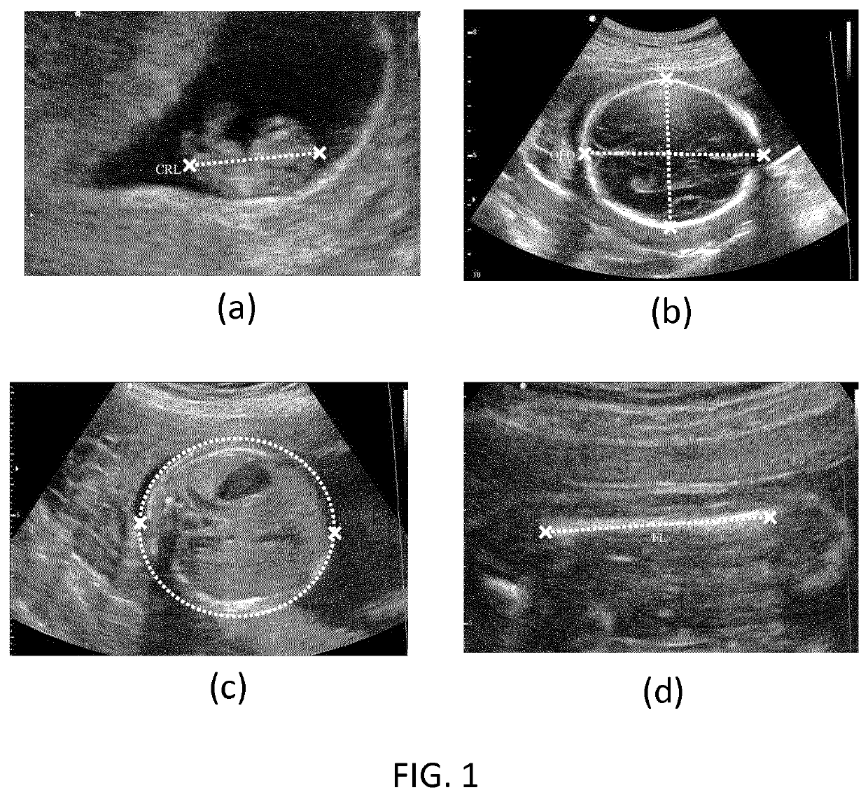
FIELD OF THE INVENTION[0001]This invention relates to a method and device for obtaining anatomical measurements from an ultrasound image.BACKGROUND OF THE INVENTION[0002]Ultrasound imaging is widely used for investigating the health and characteristics of internal anatomical structures. It may be used for instance in investigating the heart, kidneys or lungs. As part of such investigations, it is often necessary to take measurements of investigated structures, to assist in building a clinical picture.[0003]By way of example, ultrasonic imaging is routinely used during pregnancy to assess the development of a fetus in the mother's womb, for example to detect structural anomalies in the fetus. The traditional way for a clinician to acquire an image of each required view of the fetus is to manipulate an ultrasound probe while in acoustic contact with the abdomen of the mother until a desired anatomical orientation is in the plane of the 2D imaging probe. If multiple views are to be gen...

Claims


Application Information

Patent Timeline
Patent Type & Authority Applications(United States)
IPC IPC(8): A61B8/08A61B8/00G06T7/62G06T7/00
CPCA61B8/0866A61B8/463A61B8/465A61B8/469G06T2207/20101G06T7/0012G06T2207/10132G06T2207/30044G06T7/62A61B8/468A61B8/5223A61B5/1076G06T2207/30004G16H30/40G16H40/63G16H50/30G16H50/20
Inventor SCHADEWALDT, NICOLELORENZ, CRISTIANSCHMIDT-RICHBERG, ALEXANDERKLINDER, TOBIAS
Owner KONINKLJIJKE PHILIPS NV
Learn More