Catheter assembly and method for treating bifurcations

a catheter and assembly technology, applied in the field of catheter and stent delivery and deployment assemblies, can solve the problems of affecting the delivery of stents at bifurcations, and achieve the effects of improving deliverability, deliverability and manufacturability, and saving physician time and effor

Inactive Publication Date: 2008-01-10
ABBOTT CARDIOVASCULAR
View PDF99 Cites 63 Cited by
  • Summary
  • Abstract
  • Description
  • Claims
  • Application Information

AI Technical Summary

Benefits of technology

[0049]In one embodiment, the bifurcated distal section of the catheter has at least one secured portion along which the first and second branches of the distal shaft section are permanently secured together. The secured portion is located proximal to the inflatable section of the balloons, and preferably distal to the proximal end of the branched distal shaft section (and distal to the intermediate section of the shaft) so that the branched distal shaft section has an unsecured portion which is proximally adjacent to the secured portion. In a presently preferred embodiment, the two shafts are permanently secured together at least in part by a tubular outer band member such as, for example, a length of heat shrink tubing. A first band member is preferably located adjacent to the proximal-most balloon end, and a second band member is preferably located proximal thereto. For example, in a presently preferred embodiment, the second band member is located at about the half-way point along the length of the distal shaft section between the proximal end of the balloons and the distal end of the intermediate shaft section. Adhesive may additionally or alternatively be used to join the branches together. The secured distal branches of the catheter provide improved deliverability by preventing or inhibiting the tendency of the two outer members of the distal shafts to separate during advancement of the catheter within the patient's tortuous anatomy. As a result, the catheter has the deliverability and manufacturability advantages provided by the two shafts extending separately from the intermediate shaft section, in combination with the deliverability advantages provided by securing the two shafts together at one or more location between the balloons and the intermediate shaft section. Additionally, the secured distal shafts of the catheter prevent or inhibit damage to the vessel wall which can otherwise occur if the end of the stent is caused to become flared. Such flaring at the end of the stent can occur as the proximal ends of the balloons move a disadvantageously large amount relative to one another during delivery and deployment of the stent.
[0050]A joining wire slidably disposed within a branch (typically the OTW branch) of the catheter releaseably joins the distal tips of the two branches of the catheter together for advancement within the patient's anatomy. The joining wire is locked to the proximal end of the catheter assembly to keep the two distal tips together during delivery to ensure that the stent remains securely mounted on the balloons. With the stent in position for deployment within the body lumen, the joining wire is at least partially retracted to release the two branch tips. However, the ability to advance the joining wire within the branch vessel and use the joining wire to seat the stent into position within the vessel depends on the member used to lock the joining wire to the proximal end of the catheter assembly. For example, a joining wire which has a proximal end which is trimmed and secured to a connector at the proximal end of the catheter assembly must therefore be fully withdrawn and replaced with a separate guidewire for use in seating the stent into position within the vessel. In one embodiment of the invention, the joining wire also functions as a guidewire, with a proximal end slidably disposed out the proximal end of a guidewire locking mechanism releaseably securing the joining guidewire to the proximal end of the catheter. For example, in one embodiment, the guidewire locking mechanism has a collet member with a radially collapsible slotted head positioned within a proximal adapter or fitting on the proximal end of the catheter shaft. The guidewire locking mechanism saves physician time and effort by avoiding the removal and replacement of the joining wire. Additionally, the guidewire locking mechanism preferably is configured to facilitate manufacture of the catheter assembly and loading of the joining guidewire into the catheter. In one embodiment, the guidewire locking mechanism is in the locked mode with the distal end of the joining guidewire positioned distally beyond the distal end of the catheter first branch. In this configuration, the first branch and joining guidewire act as a fixed wire device, which is particularly preferred for delivering low profile devices through long, tortuous or diffusely deceased vasculature.
[0051]In one embodiment, the guidewire locking mechanism comprises a guidewire locking torque handle (“torquer”) on a proximal end section of the joining guidewire. The torquer reversibly engages the joining guidewire to provide a finger hold for manipulating the joining guidewire. Additionally, the torquer releaseably connects to a proximal end of the catheter assembly (e.g. to the proximal adapter) to thereby releasably lock the joining guidewire to the catheter. In the locked configuration, the joining guidewire is held in place relative to the catheter, and in the unlocked configuration the joining guidewire is free to slide within the catheter with the torquer connected to the joining guidewire to provide a handle for the physician facilitating the independent manipulation of the joining guidewire. As a result, the catheter assembly limits procedure time and steps, with a joining wire that functions as both a useable guidewire that can be steered, and as a joining wire that releasably joins the distal tips of the two shafts together. Although discussed primarily in terms of use with a bifurcated stent delivery catheter, the guidewire locking torque handle can be used with a variety of suitable catheters having an over-the-wire shaft design in which the guidewire is slidably disposed in the catheter guidewire lumen. Therefore, with the guidewire locking torque handle tightened down onto the guidewire and simultaneously secured to the proximal end of an over-the-wire catheter (e.g., to the proximal adapter / hub), the guidewire locking torque handle of the invention provides a stable and fixed position and relation between the guidewire and catheter, so that the catheter can be advanced or withdrawn from the body lumen while maintaining its position relative to the guidewire.
[0052]A method of delivering and implanting the stent mounted on the catheter assembly is contemplated by the present invention. The bifurcated catheter assembly of the present invention provides two separate balloons in parallel which are advanced into separate passageways of an arterial bifurcation and the balloons are inflated either simultaneously or independently (or a combination thereof) to expand and implant the stent. More specifically, and in keeping with the invention, the catheter assembly is advanced through a guiding catheter (not shown) until the distal end of the catheter assembly reaches the ostium to the coronary arteries. An Rx guide wire is advanced into the coronary arteries to a point distal of the bifurcation or target site. In a typical procedure, the Rx guide wire will already be positioned at the target site after a pre-dilatation procedure. The catheter assembly is advanced over the Rx guide wire so that the catheter distal end is just proximal to the opening to the side branch vessel. Up to this point in time, the OTW guide wire (or mandrel or joining wire) remains within the catheter assembly and within the coupler so that the long balloon and the short balloon of the catheter assembly remain adjacent to one another to provide a low profile and prevent wire wrap. As the catheter assembly is advanced to the bifurcated area, the coupler moves axially relative to the distal end of the OTW guide wire (or mandrel or joining wire) a small distance (approximately 0.5 mm up to about 5.0 mm), but not pull completely out of the coupler, making it easier for the distal end of the catheter to negotiate tortuous turns in the coronary arteries. Thus, the slight axial movement of the coupler relative to the OTW guide wire (or mandrel or joining wire) distal end allows the tips to act or move independently, thereby increasing flexibility over the tips joined rigidly and it aids in the smooth tracking of the catheter assembly over the Rx guide wire. The proximal end of the OTW guide wire is releasably attached to the proximal hub as previously described. The OTW guide wire (or mandrel or joining wire) is removed or withdrawn proximally from the coupler, thereby uncoupling the long balloon and the short balloon. Thereafter, the OTW guide wire is advanced distally into the side branch vessel so that the catheter assembly can next be advanced distally over the Rx guide wire in the main vessel and the OTW guide wire in the side branch vessel. The separation between the Rx guide wire and the OTW guide wire allows the long balloon and the short balloon to separate slightly as the catheter assembly is further advanced over the Rx guide wire and the OTW guide wire. The catheter assembly advances distally until it reaches a point where the central opening on the stent is approximately adjacent to the opening to the side branch vessel, so that the catheter assembly can no longer be advanced distally since the balloons push against the carina and are somewhat constrained by the stent. One or more high percent tungsten / radiopaque markers are placed on the distal portion of the PEBAX balloon catheter assembly to aid in positioning the stent with respect to the bifurcation or target site. Once the long and short balloons with the stent mounted thereon are positioned in the main vessel just proximal to the side branch vessel, the long balloon and the short balloon are next inflated simultaneously or independently (or a combination thereof), to expand the stent in the main vessel and the opening to the side branch vessel. The central section of the stent is expanded into contact with the opening to the side branch vessel and the central opening should substantially coincide with the opening to the side branch vessel providing a clear blood flow path through the proximal opening of the stent and through the central opening into the side branch vessel. By inflating the long balloon and the short balloon substantially simultaneously, plaque shifting is avoided and access to the side branch is better preserved.
[0053]As discussed above, as the catheter assembly is advanced through tortuous coronary arteries, over the Rx guide wire, the central opening of the stent may or may not always be perfectly aligned with the opening to the side branch vessel, and may thus be “out of phase,” and depending upon how many degrees out of phase, the stent may require repositioning or reorienting so that the central opening more closely coincides with the opening to the side branch vessel. The orientation of the central opening of the stent with respect to the opening to the side branch vessel can range anywhere from a few degrees to 180°. If the central opening of the stent is more than 90° out of phase with respect to the opening to the side branch vessel, it may be difficult to position the radiopaque marker, and thus the linear or longitudinal position of the stent. When the central opening is in the out of phase position, the stent of the invention still can be implanted and the central opening will expand into the opening of the side branch vessel and provide adequate coverage. In cases where the system is more than 90° out of phase, the Rx and OTW guide wires will be crossed causing a distal torque to be applied to help the system to rotate in phase. In the event rotation does not occur, the system can be safely deployed with adequate coverage and support as long as the radiopaque markers located on the distal end of the catheter reach the proper positioning as can be detected under fluoroscopy. The unique and novel design of the catheter assembly and the stent of the present invention minimizes the misalignment so that the central opening of the stent generally aligns with the opening to the side branch vessel, and is capable of stenting the opening to the side branch vessel even if the central opening is out of phase from the opening of the side branch vessel.
[0054]One aspect of the invention is directed to a method of delivering a stent to a patient's bifurcated blood vessel, generally comprising introducing and advancing within a patient blood vessel a stent delivery balloon catheter having a polymeric radiopaque distal tip marker secured to the first branch (e.g., side branch), and fluoroscopically imaging the polymeric radiopaque distal tip marker to determine the alignment of the first branch balloon relative to an opening of a side branch of the patient's blood vessel. Under fluoroscopy, the image of the polymeric radiopaque distal tip marker facilitates adjusting the alignment of the first branch balloon relative to the side branch opening of the blood vessel both before and after the two distal tips of the catheter are uncoupled. It also facilitates placement of a wire in the side branch vessel after the joining mandrel is removed by making visible the tip where the wire will exit the catheter.

Problems solved by technology

Repair of vessels that are diseased at a bifurcation is particularly challenging since the stent must be precisely positioned, provide adequate coverage of the disease, provide access to any diseased area located distally to the bifurcation, and maintain vessel patency in order to allow adequate blood flow to reach the myocardium.
Where the stent provides coverage to the vessel at the diseased portion, yet extends into the vessel lumen at the bifurcation, the diseased area is treated, but blood flow may be compromised in other portions of the bifurcation.
Unapposed stent elements may promote lumen compromise during neointimal formation and healing, producing restenosis and requiring further procedures.
Moreover, by extending into the vessel lumen at the bifurcation, the stent may block access to further interventional procedures.
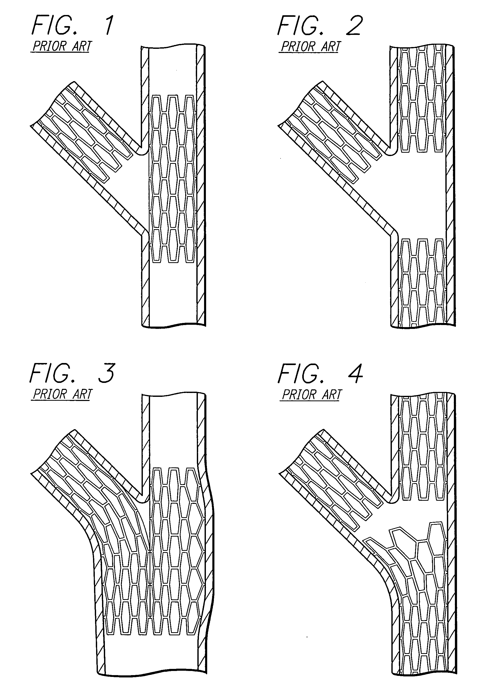
Conventional stents are designed to repair areas of blood vessels that are removed from bifurcations and, therefore, are associated with a variety of problems when attempting to use them to treat lesions at a bifurcation.
The drawback with this approach is that there is no way to determine or guarantee that the main-vessel stent struts are properly oriented with respect to the side branch or that an appropriate stent cell has been selected by the wire for dilatation.
The aperture created often does not provide a clear opening and creates a major distortion in the surrounding stent struts.
A further drawback with this approach is that it is difficult to determine if the stent struts in the stented vessel have been properly oriented and spread apart to provide a clear opening for stenting the jailed vessel.
This technique also causes stent deformation to occur in the area adjacent to the carina, pulling the stent away from the vessel wall and partially obstructing flow in the originally non-jailed vessel.
Deforming the stent struts to regain access into the previously jailed vessel is also a complicated and time consuming procedure associated with attendant risks to the patient and is typically performed only if considered an absolute necessity.
The risks of procedural complications during this subsequent deformation are considerably higher than stenting in normal vessels.
The inability to place a guide wire through the jailed lumen in a timely fashion could restrict blood supply and begin to precipitate symptoms of angina or even cardiac arrest.
In addition, disturbed hemodynamics and subsequent thrombus formation at the jailed site could further compromise blood flow into the side branch.
This procedure is also associated with the same issues and risks previously described when stenting only one vessel and deforming the struts through the jailed vessel.
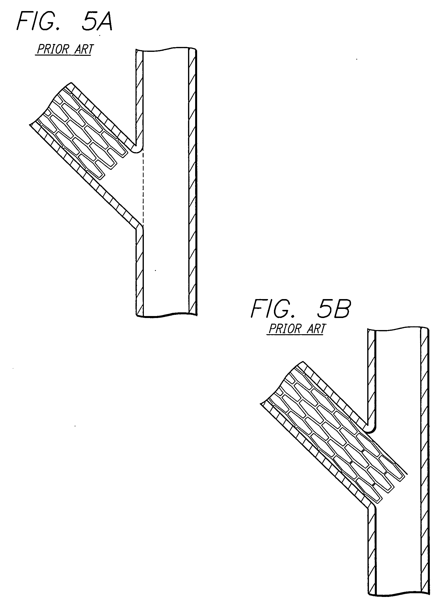
In addition, since a conventional stent generally terminates at right angles to its longitudinal axis, the use of conventional stents to treat the origin of the previously jailed vessel (typically the side branch vessel) may result in blocking blood flow of the originally non-jailed vessel (typically the parent vessel) or fail to provide adequate coverage of the disease in the previously jailed vessel (typically a side branch vessel).
Such a position of the conventional stent results in a bifurcation that does not provide full coverage or has a gap on the proximal side (the origin of the side branch) of the vessel and is thus not completely supported.
One of the drawbacks of this approach is that the orientation of the stent elements protruding from the side branch vessel into the main vessel is completely random.
In addition excessive metal coverage exists from overlapping strut elements in the parent vessel proximal to the carina area.
When dilating the main vessel the stent struts are randomly stretched, thereby leaving the possibility of restricted access, incomplete lumen dilatation, and major stent distortion.
All of the foregoing stent deployment assemblies suffer from the same problems and limitations.
Typically, there are uncovered surface segments or overlapped struts on the main vessel and side branch vessels between the stented segments, or there is excessive coverage in the parent vessel proximal to the bifurcation.
An uncovered flap or fold in the intima or plaque will invite a “snowplow” effect, representing a substantial risk for sub-acute thrombosis, and the increased risk of the development of restenosis.
Further, where portions of the stent are left unapposed within the lumen, the risk for subacute thrombosis or the development of restenosis again is increased.
The prior art stents and delivery assemblies for treating bifurcations are difficult to use and deliver making successful placement challenging.
Further, even where placement has been successful, the side branch vessel can be “jailed” or covered so that there is impaired access to the stented area for subsequent intervention.
In addition to problems encountered in treating disease involving bifurcations for vessel origins, difficulty is also encountered in treating disease confined to a vessel segment but extending very close to a distal branch point or bifurcation which is not diseased and does not require treatment.
In such circumstances, very precise placement of a stent covering the diseased segment, but not extending into or obstructing the side branch, may be difficult or impossible.

Method used


Image

  • Catheter assembly and method for treating bifurcations
  • Catheter assembly and method for treating bifurcations
  • Catheter assembly and method for treating bifurcations

Examples


Embodiment Construction

[0120]The present invention includes a stent and stent delivery catheter assembly and method for treating bifurcations in, for example, the coronary arteries, veins, peripheral vessels and other body lumens. Prior art attempts at implanting intravascular stents in a bifurcation have proved less than satisfactory. For example, FIGS. 1-4 depict prior art devices which include multiple stents being implanted in both the main vessel and a side branch vessel. In FIG. 1, a prior art “T” stent is implanted such that a first stent is implanted in the side branch near the origin of the bifurcation, and a second stent is implanted in the main vessel. With this approach, portions of the side branch vessel are left uncovered, and blood flow to the side branch vessel must necessarily pass through the main vessel stent, causing possible obstructions or thrombosis.

[0121]Referring to FIG. 2, three prior art stents are required to stent the bifurcation which leaves a large gap in vessel coverage bet...


PUM


Abstract

An improved stent design and stent delivery catheter assembly for repairing a main vessel and a side branch vessel forming a bifurcation. The stent includes rings aligned along a common longitudinal axis and connected by links, where the stent has one or more portals for aligning with and partially expanding into the opening to the side branch vessel. The stent is implanted at a bifurcation so that the main stent section is in the main vessel, and the portal section covers at least a portion of the opening to the side branch vessel. A second stent can be implanted in the side branch vessel and abut the expanded central section to provide full coverage of the bifurcated area in the main vessel and the side branch vessel. Radiopaque markers on the stent and on the tip of the delivery catheter assist in aligning the portal section with the opening to the side branch vessel.

Description

[0001]This application is a continuation of currently pending U.S. patent application Ser. No. 11 / 483,300 filed Jul. 7, 2006.BACKGROUND OF THE INVENTION[0002]The invention relates to stents and stent delivery and deployment assemblies for use at a bifurcation and, more particularly, one or more stents for repairing bifurcations, blood vessels that are diseased, and a method and apparatus for delivery and implantation of the stents.[0003]Stents conventionally repair blood vessels that are diseased. Stents are generally hollow and cylindrical in shape and have terminal ends that are generally perpendicular to their longitudinal axis. In use, the conventional stent is positioned at the diseased area of a vessel and, after deployment, the stent provides an unobstructed pathway for blood flow.[0004]Repair of vessels that are diseased at a bifurcation is particularly challenging since the stent must be precisely positioned, provide adequate coverage of the disease, provide access to any d...

Claims


Application Information

Patent Timeline
Patent Type & Authority Applications(United States)
IPC IPC(8): A61F2/84A61F2/06
CPCA61F2/856A61F2/91A61F2/915A61F2/954A61F2250/006A61F2002/067A61F2002/91525A61F2002/91575A61F2/958
Inventor TA, DIEM UYENWU, CAROLINEHEARN, BRENNA K.COX, DANIEL L.BARBOD, LEONARD D.DELGADO, JESSIEPACETTI, STEPHEN DIRKHATTEN, THOMAS RAYWROLSTAD, DAVID K.ARMSTRONG, KENNETH KAYKENT, DARRIN J.
Owner ABBOTT CARDIOVASCULAR
Why Eureka
Try Eureka