Device and Method for Preparing Sample Material

a sample material and device technology, applied in the field of sample removal devices, can solve the problems of high development cost, loss of advantage of almost completely automatic processing, and danger of contamination or decomposition of fragile sample materials, so as to reduce the number of manual steps, reduce the number of errors, and the effect of high degree of sensitivity

Pending Publication Date: 2021-04-22
ROBERT BOSCH GMBH
View PDF0 Cites 0 Cited by
  • Summary
  • Abstract
  • Description
  • Claims
  • Application Information

AI Technical Summary

Benefits of technology

[0003]The device for preparing sample material is advantageously configured as a rotary device with which, by means of a rotation movement, a defined quantity of liquid can be drawn into a sample-receiving space for the sample material or can be expelled from the sample-receiving space for the sample material. The sample-receiving space preferably has a microfluidic volume, in particular a liquid volume, of one to one thousand microliters, preferably between ten and one hundred microliters. The sample material is, for example, animal, human and / or plant tissue. The sample material is removed, for example, with a suitable sample removal device. The sample removal device is, for example, a biopsy needle or a biodetector with a functional or functionalized surface. The functional or functionalized surface advantageously serves to isolate molecules or cells from the human body. Cells, in particular tumor cells, circulating in the blood stream can be removed from a patient with the biopsy needle. The functional or functionalized part of the biopsy needle is advantageously coated such that either cell-free DNA or cells of epithelial origin, expressing a defined surface protein such as EpCAM, come into contact with the needle surface and are bound by antibodies present there, such as anti-Ep-CAM. The abbreviation Ep-CAM stands for epithelial cell adhesion molecule. In one use, the biopsy needle or the biodetector is introduced for example for thirty minutes into the brachial vein of a patient, removed and washed. A physician then determines the number of fixed cells and / or determines the mutation state of the cells. In a microfluidic system, small sample quantities can be analyzed with a high degree of sensitivity. Automation, miniaturization and parallelization additionally permit a reduction in the number of manual steps and, consequently, fewer errors caused by such steps. Prior to a microfluidic analysis process, however, the macroscopic sample must first of all be transferred into the microscopic or fluidic fluid environment.

Problems solved by technology

There is also the danger of contamination or decomposition of the fragile sample material as a result of time-intensive steps.
To this end, the microfluidic analysis unit would often have to be newly adapted, which is associated with high development costs.
Otherwise, the advantage of the almost completely automatic processing is lost.

Method used


Image

  • Device and Method for Preparing Sample Material
  • Device and Method for Preparing Sample Material
  • Device and Method for Preparing Sample Material

Examples


Embodiment Construction

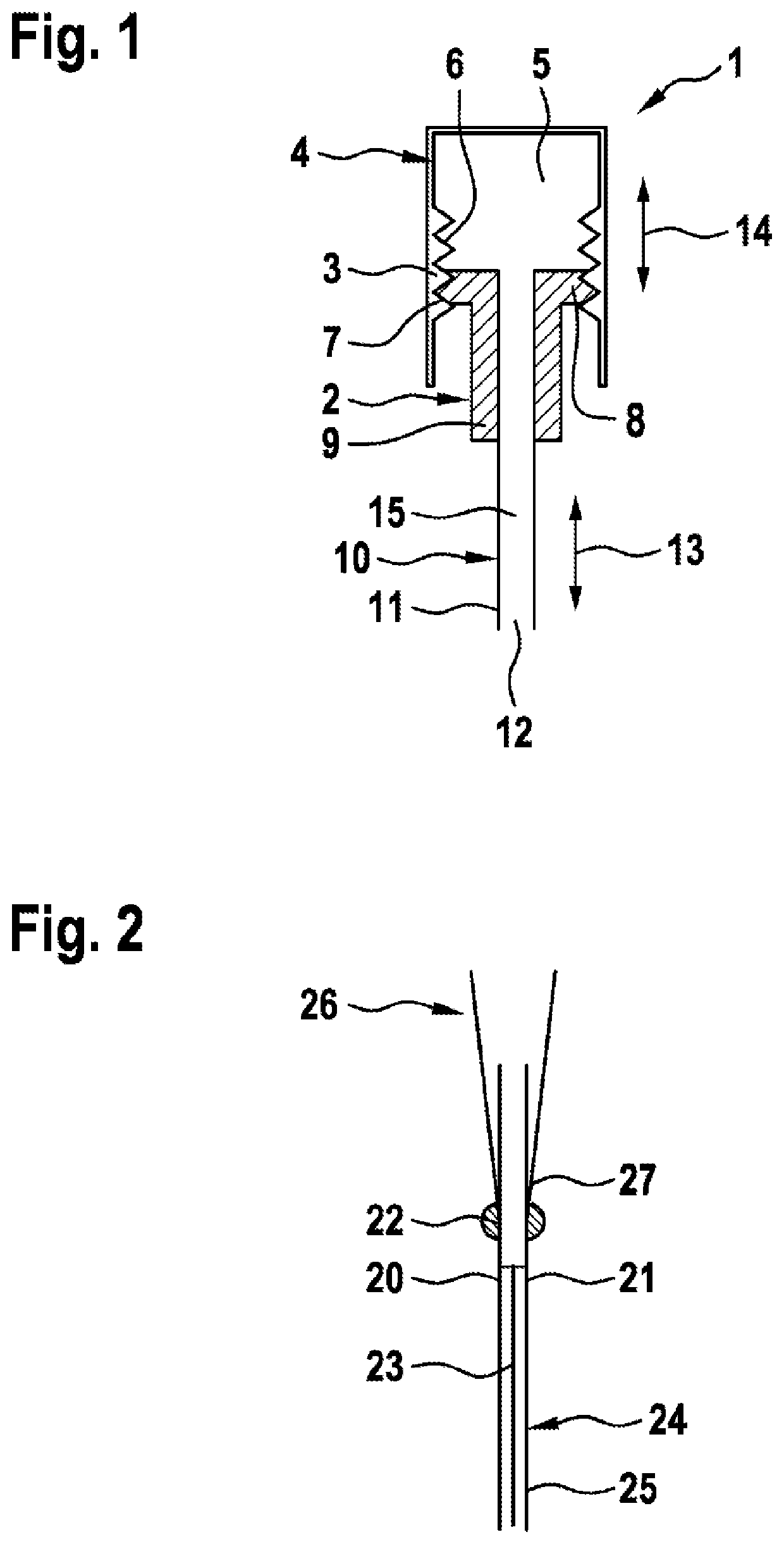
[0058]FIG. 1 shows schematically, in longitudinal section, a device 1 configured as a rotary device for preparing sample material 23, 33 (in FIGS. 2, 3, 5 to 8, 12, 13).

[0059]The rotary device 1 comprises a rotary body 2 which is rotatable in a hollow body 4 with the aid of a thread 3. The hollow body 4 has the shape of a straight circular cylinder, which is closed at its upper end in FIG. 1. The hollow body 4 delimits a liquid-receiving space 5.

[0060]The thread 3 comprises an inner thread portion 6 in the hollow body 4. An outer thread portion 7 of the thread 3 engages in the inner thread portion 6. The outer thread portion 7 is formed on a collar 8 of the rotary body 2. The collar 8 is angled away from a main body 9 of the rotary body 2.

[0061]The main body 9 of the rotary body 2 comprises a central through-hole, which leads into a tube body 10. The tube body 10 is configured, for example, as a capillary 11 with an open end 12 at the bottom. The capillary 11 delimits on the inside ...


PUM

PropertyMeasurementUnit
volumeaaaaaaaaaa
volumeaaaaaaaaaa
volumeaaaaaaaaaa

Abstract

A device for preparing sample material is designed as a rotary device by means of which a defined quantity of liquid can be drawn in into a sample receiving chamber for the sample material or can be expelled from the sample receiving chamber for the sample material, by means of a rotary motion.

Description

[0001]The invention relates to a device and a method for preparing sample material. The invention further relates to a microfluidic system having such a device.PRIOR ART[0002]The German laid-open specification DE 10 2005 050 347 A1 discloses a sample removal device, in particular a biopsy needle, composed of a hollow needle, having a distal opening with a peripheral edge, and of a stylet which is guided displaceably in the hollow needle and has a tip and a length such that the tip can protrude from the distal opening of the hollow needle. The sample removal device is configured, for example, as a fine needle biopsy device. The sample removal device or fine needle biopsy device is used to remove animal, human and / or plant tissue. In cases of suspected disease, fine needle biopsies are performed to remove tissue material or cells from the lungs, thyroid gland or prostate, for example. This sample material is traditionally placed onto a slide and assessed by a pathologist. The assessme...

Claims


Application Information

Patent Timeline
Patent Type & Authority Applications(United States)
IPC IPC(8): B01L3/00C12M1/26
CPCB01L3/502715C12M33/04B01L2200/027B01L2400/0487B01L2300/044B01L2300/0672B01L2300/0681B01L2200/0689A61M39/10A61B10/0233G01N1/28
Inventor LAERMER, FRANZHOFFMANN, JOCHENFRANK, TINOKALLENBERGER, PAUL
Owner ROBERT BOSCH GMBH
Why Eureka
Try Eureka