System and method for imaging, segmentation, temporal and spatial tracking, and analysis of visible and infrared images of ocular surface and eye adnexa

a technology of visible and infrared images and system and methods, applied in image data processing, image enhancement, medical science, etc., can solve the problems of inability to segment images, inability to track eye movements, and lack of resolution

Pending Publication Date: 2021-10-21
ZARE BIDAKI EHSAN +2
View PDF0 Cites 5 Cited by
  • Summary
  • Abstract
  • Description
  • Claims
  • Application Information

AI Technical Summary

Benefits of technology

[0016]The present disclosure relates to a system and method for imaging and analysis of ocular disease and studies of the eye. More generally, the present system and method provides an improved system and method for automatically and non-invasively imaging the ocular surface and adnexa tissues using infrared (IR) thermal cameras and visible light cameras synchronously. In an embodiment, the present system and method segments the images produced to identify specific ocular structures and measures ocular surface temperature (OST) within segmented areas by tracking the OST precisely, including by monitoring eye tracking and eye blinking during measurement to remove artefacts and maintain synchronicity. Temporal and spatial changes in the IR and visible images are tracked over time, and diagnostic indicators for ocular disease diagnosis and study of the eye are produced.

Problems solved by technology

They lack resolution in the IR image, and the image cannot be segmented, eye movement cannot be tracked, and artefacts from eyelid blinking cannot be removed.
However, it has significant limitations.
These limitations make such a system impractical and infeasible to use for imaging and analysis of ocular surfaces for ocular disease and studies of the eye in practical scenarios, especially real-time, high-speed scenarios, given the time consuming, manual nature of such a configuration and is prone to error.
In this configuration, the corneal boundary of the subject's eye could be identified with improved accuracy, but the two cameras were not synchronized and so is impractical and infeasible to use for imaging and analysis of ocular surfaces for ocular disease and studies of the eye in practical scenarios, especially real-time, high-speed scenarios, given the need for manual processing of collected data and is also prone to error.
Each of the above references has various limitations which may inhibit full realization of imaging and analysis techniques.

Method used


Image

  • System and method for imaging, segmentation, temporal and spatial tracking, and analysis of visible and infrared images of ocular surface and eye adnexa
  • System and method for imaging, segmentation, temporal and spatial tracking, and analysis of visible and infrared images of ocular surface and eye adnexa
  • System and method for imaging, segmentation, temporal and spatial tracking, and analysis of visible and infrared images of ocular surface and eye adnexa

Examples


Embodiment Construction

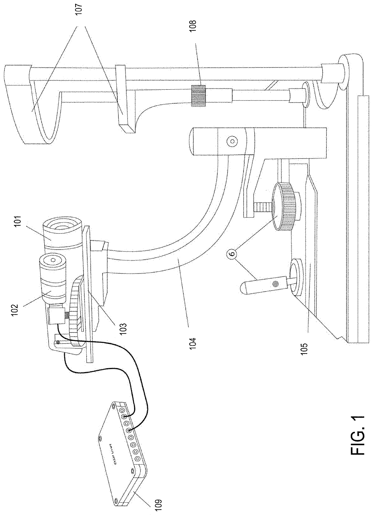
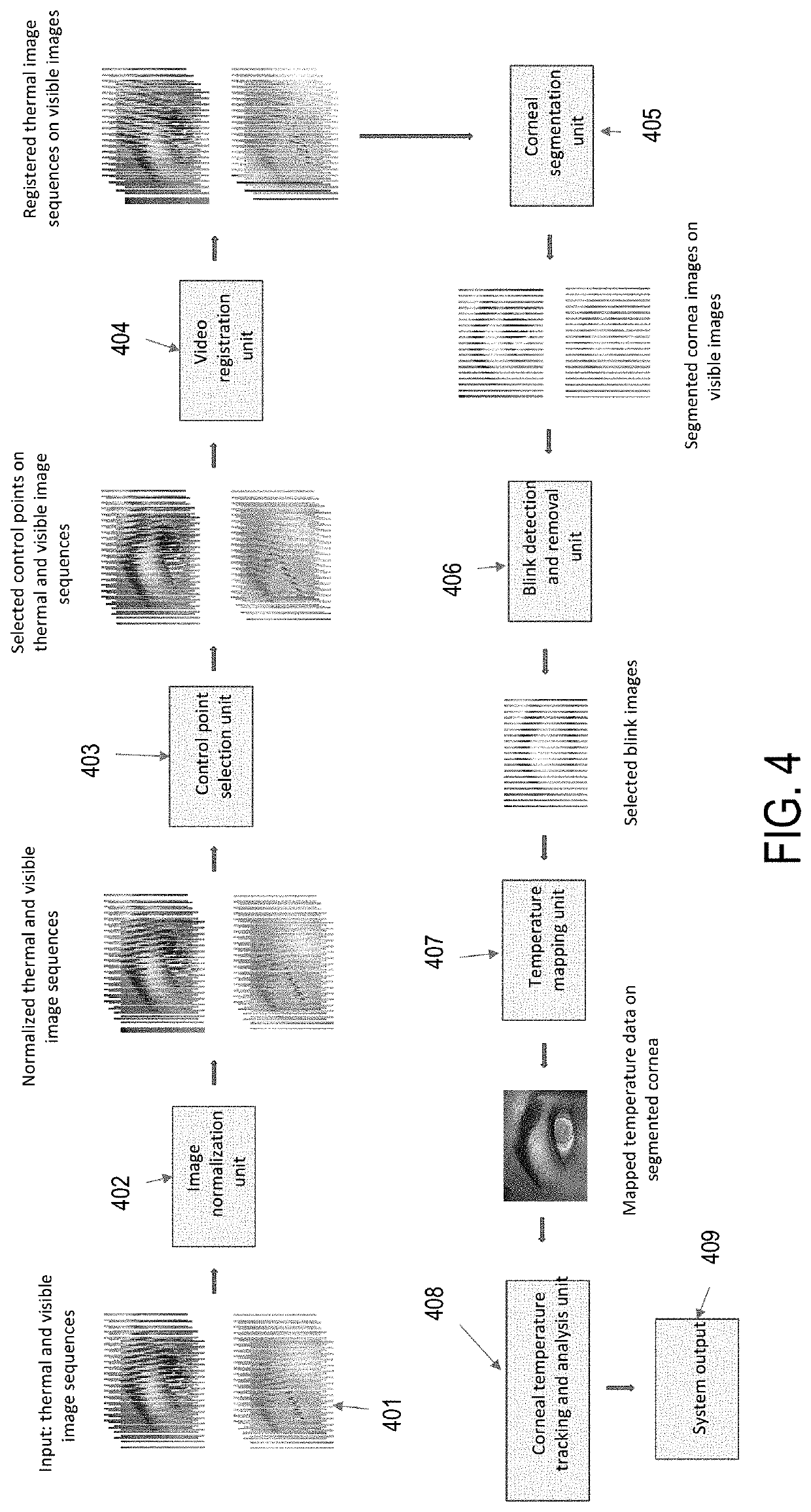
[0038]As noted above, the present disclosure relates to a system and method for imaging and analysis of ocular disease and studies of the eye. More generally, the present system and method provides an improved system and method for automatically and non-invasively imaging the ocular surface and adnexa tissues using infrared (IR) thermal cameras and visible light cameras synchronously. In an embodiment, the present system and method segments the images produced to identify specific ocular structures and measures ocular surface temperature (OST) within segmented areas by tracking the OST precisely, including by monitoring eye tracking and eye blinking during measurement to remove artefacts and maintain synchronicity. Temporal and spatial changes in the IR and visible images are tracked over time, and diagnostic indicators for ocular disease diagnosis and study of the eye are produced.

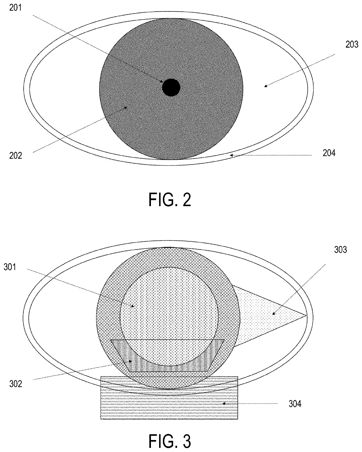
[0039]A key requirement for ocular surface thermography is the ability to locate the corneal area in t...


PUM


Abstract

An automatic system and method for non-invasive imaging and identification of specific ocular structures of the eye and adnexa tissues by synchronous segmentation of visual and infrared images; that can produce spatial temperature profiles within each segmented area of the eye and adnexa; that can track eye and head movement and eye-blinks during the period of measurement to remove artefacts and maintain synchronicity; that can track ocular surface and eye adnexa temperature profiles over time; that can assist in diagnosis of eye disease; that can produce diagnostic indicators for ocular disease diagnosis and study of the eye. The system comprises infrared and visible light cameras for imaging the ocular structures, and a digital signal processing unit for processing the acquired infrared and visible images to output segmentations of the images for identification of different areas of the eye surface, including pupil, cornea, conjunctiva, and eyelids. The system further captures synchronous infrared and visible images from each segmented area of the ocular surface over the time of measurement. A digital signal processing unit processes and analyzes the infrared and visible images to generate descriptive outputs on temporal and spatial changes in the infrared and visible images over the time of measurement, as well as produce diagnostic indicators for ocular disease diagnosis and study of the eye.

Description

CROSS-REFERENCE TO RELATED APPLICATIONS[0001]This application claims priority from U.S. Provisional Application No. 63 / 012,965 filed on Apr. 21, 2020, which is incorporated by reference herein in its entirely.FIELD OF THE INVENTION[0002]The present disclosure relates to a system and method for imaging and analysis of ocular surfaces for ocular diseases and studies of the eye.BACKGROUND[0003]Body temperature reflects physiological information about human health. It can be assessed using an invasive (contact) method, e.g. thermal expansion of a liquid, or a non-invasive (non-contact) method, e.g. IR imaging (thermography). IR imaging has many advantages over contact methods: it is non-invasive and so does not alter the target tissue structure or stability which would alter the temperature profile, can be obtained in real-time, can provide continuous data over the time period of measurement, can provide data over a large surface area, and is very accurate. Clinically, IR imaging is use...

Claims


Application Information

Patent Timeline
Patent Type & Authority Applications(United States)
IPC IPC(8): A61B5/01G06T5/50G06T7/11G06T7/00A61B3/14A61B5/11A61B3/113A61B3/10
CPCA61B5/015G06T5/50G06T7/11G06T7/0016G06T2207/30041A61B5/1128A61B3/113A61B3/101G06T2207/10048A61B3/14A61B5/6821A61B5/0077A61B3/0025A61B3/18G06T2207/10024G06T7/174G06T2207/10016
Inventor ZARE BIDAKI, EHSANMURPHY, PAUL J.WONG, ALEXANDER SHEUNG LAI
Owner ZARE BIDAKI EHSAN
Why Eureka
Try Eureka