Supercharge Your Innovation With Domain-Expert AI Agents!

Method for inducing differentiation of corneal epithelial cells from pluripotent stem cells

a technology of stem cells and cornea, which is applied in the field of inducing the differentiation of corneal epithelial cells from pluripotent stem cells, can solve the problems of rejection reaction, many limitations in research and use of stem cells, and limited applications of es cells, so as to achieve high safety and clinical application. suited

Pending Publication Date: 2021-12-02
OSAKA UNIV
View PDF0 Cites 0 Cited by
  • Summary
  • Abstract
  • Description
  • Claims
  • Application Information

AI Technical Summary

Benefits of technology

The patent describes a method to create corneal epithelial cells from human iPS cells. These cells have specific markers and are safe for clinical application as they do not use animal-derived materials. Additionally, the method can also be used to create other eye cells such as conjunctival epithelial cells, retinal cells, and lacrimal gland cells.

Problems solved by technology

However, the research and use of the stem cells have many limitations due to the ethical problem of the destruction of fertilized ova in their establishment, and there is also a problem of rejection reaction since ES cells derived from a patient who is to undergo transplantation cannot be established.
Thus, in the present situation, diseases to which ES cells are applicable are limited and their clinical usefulness is still not evident.
The inventors have reported differentiation into corneal epithelium among stratified epithelia (Patent Literatures 2 to 3 and Non Patent Literature 3); however, the inability to be made into sheet, the inability to induce corneal epithelial progenitor cells having a high growth capacity, and the use of feeder cells have been problems in clinical application.

Method used


Image

  • Method for inducing differentiation of corneal epithelial cells from pluripotent stem cells
  • Method for inducing differentiation of corneal epithelial cells from pluripotent stem cells
  • Method for inducing differentiation of corneal epithelial cells from pluripotent stem cells

Examples


example 1

of Differentiation from Human iPS Cell into Ocular Surface Epithelial / Corneal Epithelial Cell

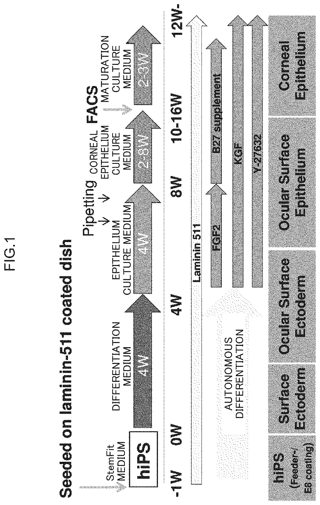
[0146]From human iPS cells, ocular surface epithelial / corneal epithelial cells were differentiated in a serum-free medium without using feeder cells. The scheme of the method for differentiation induction is shown in FIG. 1.

[0147]The 201B7 cell line (an iPS cell line prepared by introducing Oct3 / 4, OX2, c-Myc, and Klf4 into fibroblasts) was used as human iPS cells, and the cells were seeded at a seeding density of 200 to 1,500 cells / cm2, 300 to 1,000 cells / cm2, or 450 to 700 cells / cm2 in StemFit (from Ajinomoto Co., Inc.) on a 6-well plate coated with laminin 511E8 fragment, and cultured for 7 to 13 days.

[0148]The medium was replaced with a differentiation medium (GMEM containing 10% KSR, 1 mM sodium pyruvate, 0.1 mM non-essential amino acids, and 55 monothioglycerol), and then replaced with the fresh differentiation medium once every 2 to 3 days to perform culture for about 4 weeks.

[0149]Th...

example 2

of Differentiation of Corneal Epithelial Progenitor Cell from Human iPS Cell

[0156]The colonies having the form of concentric circles, formed by the autonomous differentiation (4 weeks) of human iPS cells according to Example 1 were each isolated by replacing the medium with a differentiation medium for epithelium containing a growth factor at or after the 4th week and removing non-epithelial cells by pipetting from ocular surface epithelial stem cells appearing in the 3rd zone. In addition, at or after the 8th week, the medium was replaced with a corneal epithelium culture medium, and differentiation into corneal epithelial progenitor cells was induced.

[0157]At about the 12th week of differentiation induction, FACS was carried out using cell surface markers, such as TRA-1-60, SSEA4 and ITGB4 (FIG. 8). It was identified that the corneal epithelial progenitor cells could be isolated based on the expression of TRA-1-60, SSEA4, and ITGB4 and were selectively present in ITGB4-positive an...

example 3

tation of Human iPS Cell-Derived Corneal Epithelial Cell Sheet onto Rabbit Eye

[0160]The epithelial cell layer on the whole circumference of the rabbit corneal limbus was removed by corneal abscission to prepare a rabbit corneal epithelial stem cell deficiency model. Then, a human iPS cell-derived corneal epithelial cell sheet recovered in sheet form was transplanted.

[0161](1) Ocular Surface Observation Image

[0162]At the 7th day after transplantation, corneal transparency was kept in a sheet transplantation group, and fluorescein staining showed that barrier function had been recovered. On the other hand, a large part of the control eye was stained by fluorescein and remained in a state of barrier dysfunction (FIG. 12 middle).

[0163](2) Human iPS Cell-Derived Corneal Epithelial Cell Sheet after Transplantation

[0164]Each rabbit was euthanized on postoperative day 7, and the eyeball was excised and then chemically fixed with a 10% neutral buffered formalin solution. Then, corneal tissue...


PUM


Abstract

The present invention relates to a method for inducing the differentiation of corneal epithelial cells from pluripotent stem cells. More specifically, the present invention relates to a method for autonomously differentiating pluripotent stem cells, such as human iPS cells, into ectodermal cell lineage in a serum-free medium without using feeder cells and inducing the differentiation of the resultant ocular surface ectodermal lineage cells into corneal epithelial cells.

Description

RELATED APPLICATIONS[0001]The present application is a Continuation of U.S. application Ser. No. 15 / 542,200, which is the U.S. National Stage of PCT / JP2016 / 050784, filed Jan. 13, 2016, which claims priority to Japanese Patent Application No. 2015-006074, filed on Jan. 15, 2015, the contents of which are incorporated herein by reference.TECHNICAL FIELD[0002]The present invention relates to a method for inducing the differentiation of corneal epithelial cells from pluripotent stem cells. More specifically, the present invention relates to a method for autonomously differentiating pluripotent stem cells (particularly, induced pluripotent stem cells) into ectodermal cell lineage in a serum-free medium without using feeder cells and inducing the differentiation of the resultant ocular surface ectodermal lineage cells into corneal epithelial cells.Background Art[0003]In recent years, work has been proceeding on regenerative medicine (cell medicine) for supplementing damaged tissues and or...

Claims


Application Information

Patent Timeline
Patent Type & Authority Applications(United States)
IPC IPC(8): C12N5/079C12N5/10C12N5/074
CPCC12N5/0621C12N5/10C12N5/0607C12N2506/45C12N2501/117C12N2501/727C12N2500/90C12N2501/115C12N2501/11C12N2501/15C12N2500/25A61K35/30A61K35/545C12N2501/16C12N2501/155
Inventor NISHIDAHAYASHI, RYUHEIISHIKAWA, YUKI
Owner OSAKA UNIV
Features
  • R&D
  • Intellectual Property
  • Life Sciences
  • Materials
  • Tech Scout
  • Unparalleled Data Quality
  • Higher Quality Content
  • 60% Fewer Hallucinations
Learn More