Non-invasive method and system for measuring motion characteristics of myocardial tissue

a motion characteristic and non-invasive technology, applied in the field of biological tissue technology, can solve the problems of heart failure, ventricular hypertrophy and even heart failure, and cannot explain the mechanical properties of the tissues, and achieve the effect of reducing costs

Pending Publication Date: 2022-06-16
MSHEAF HEALTH MANAGEMENT TECH LTD
View PDF0 Cites 0 Cited by
  • Summary
  • Abstract
  • Description
  • Claims
  • Application Information

AI Technical Summary

Benefits of technology

[0024]Compared with the prior art, the present invention relates to a new technology for detecting the contraction and relaxation of the myocardial tissue at the cellular level, and has the advantage that the present invention provides a continuous and non-invasive method with high-sampling rate to measure the motion of the myocardial tissue at an overall cellar level, so as to detect even more subtle abnormal changes in the myocardial cells. In addition, the present invention avoids the traditional technology of using imaging results for analysis, and provides a faster but standard measurement method with lower cost.

Problems solved by technology

Long-term hypertension will eventually lead to ventricular hypertrophy and even heart failure.
Ischemia, mainly caused by coronary artery stenosis, will eventually trigger heart attacks, followed by myocardial infarctions, and lead to a heart failure.
But these parameters do not explain the mechanical properties of the tissues.
On the one hand, currently, there has been no non-invasive measurement method for the health state of the myocardial tissue at the cellular level.
For example, the MM imaging method is very expensive.
Although ultrasonic imaging is less expensive, it is still affected by many aspects.
First, ultrasonic imaging cannot be performed continuously or for a long time.
Second, the resolution of ultrasonic imaging results is not high, and the results of ultrasonic imaging also depend on patients.
The imaging results will vary due to the lack of standardized operations, and the cost of ultrasonic imaging is still high.

Method used


Image

  • Non-invasive method and system for measuring motion characteristics of myocardial tissue
  • Non-invasive method and system for measuring motion characteristics of myocardial tissue
  • Non-invasive method and system for measuring motion characteristics of myocardial tissue

Examples


Embodiment Construction

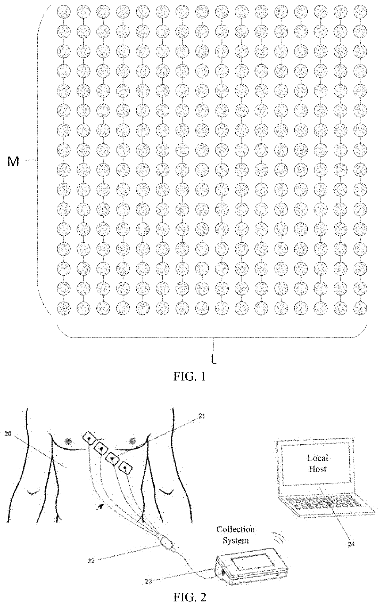
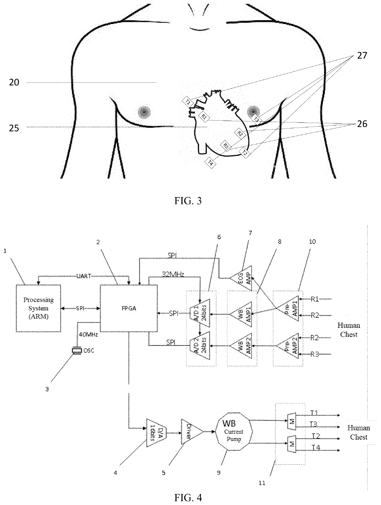
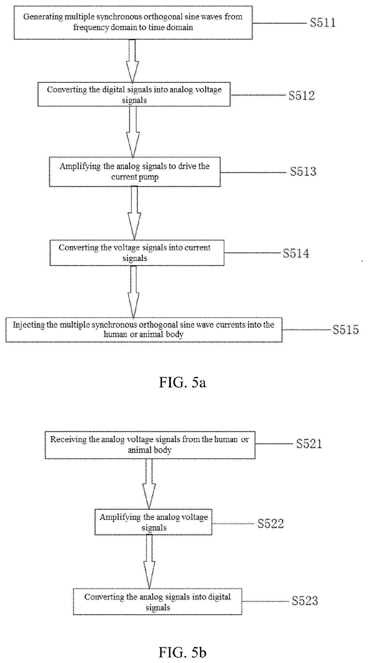
[0046]The embodiments of the present invention will now be described in further detail with reference to the drawings. These drawings are all simplified schematic diagrams, which merely illustrate the basic structure of the present invention in a schematic manner, so they only show the constitution relevant to the present invention.

[0047]The present invention relates to a non-invasive technology for detecting the electrical properties of tissues in an organism, such as resistances and capacitances of the tissues and their change patterns. Its goal is to capture changes in body fluid, blood flow and cardiovascular circulatory tissues, to monitor the health state of the organism, to measure and verify the elasticity of the cardiovascular system, and to detect information for non-therapeutic purposes.

[0048]In the embodiments provided by the present invention, heart cells are considered to be equipotential. Therefore, the cell size can be estimated by capacitance measurement. When the h...


PUM


Abstract

A non-invasive method for measuring the motion characteristics of myocardial tissue includes transmitting a plurality of generated synchronous orthogonal, phase controllable and adjustable alternating currents with different frequencies to an organism so as to generate a plurality of synchronous periodic AC voltage signals with different frequencies; receiving the periodic AC voltage signals modulated by changes in the organism's heart tissue to obtain the organism's frequency responses; calculating resistances and capacitances of the heart tissue according to the frequency responses; estimating motion characteristics of myocardial tissue according to the resistances and the capacitances. By means of introducing the average longitudinal length of the myocardial cells, and calculating changes in the average longitudinal length of the myocardial cells according to the capacitances, the overall longitudinal elasticity of the heart is described.

Description

TECHNICAL FIELD[0001]The present invention relates to a technology for measurement of biological tissues, in particular to a non-invasive method and system for measuring the motion characteristics of a myocardial tissue.BACKGROUND[0002]The basic function of a heart is to pump blood, circulate the blood in an organism, and provide oxygen and nutrients to tissues. Therefore, the measurement of cardiac dynamics parameters is of extremely important significance in the medical field. Myocardial cells have structural characteristics indicating that they pertain to an elastic tissue. Therefore, the motion of the myocardial tissue, especially elasticity thereof, should be the main measurement target. At present, the stress-strain relationship of the myocardial tissue has been extensively studied, and its related applications are mainly realized through ultrasonic imaging systems.[0003]A heart has four chambers, including two atria and two ventricles. Under normal circumstances, the right at...

Claims


Application Information

Patent Timeline
Patent Type & Authority Applications(United States)
IPC IPC(8): A61B5/0245A61B5/00
CPCA61B5/0245A61B5/7257A61B5/7475A61B5/7285A61B5/0006A61B5/0531A61B5/1126
Inventor WANG, LINGYI, CHENGHE, BIXIAXIE, PENG
Owner MSHEAF HEALTH MANAGEMENT TECH LTD
Why Eureka
Try Eureka