Systems and Methods for Automated Image Analysis

an image analysis and automated technology, applied in the field of systems and methods for automated image analysis, can solve the problems of large amount of resources, large human interaction in imaging systems, time-consuming and time-consuming processes,

Pending Publication Date: 2022-06-30
RGT UNIV OF CALIFORNIA
View PDF0 Cites 2 Cited by
  • Summary
  • Abstract
  • Description
  • Claims
  • Application Information

AI Technical Summary

Problems solved by technology

While medical imaging provides an invaluable resource, it also consumes extensive resources.
Furthermore, imaging systems require extensive human interaction to setup and operate, and then to analyze the images and make clinical decisions.
This process can be time-consuming and prone to observer variabilit...

Method used


Image

  • Systems and Methods for Automated Image Analysis
  • Systems and Methods for Automated Image Analysis
  • Systems and Methods for Automated Image Analysis

Examples


Embodiment Construction

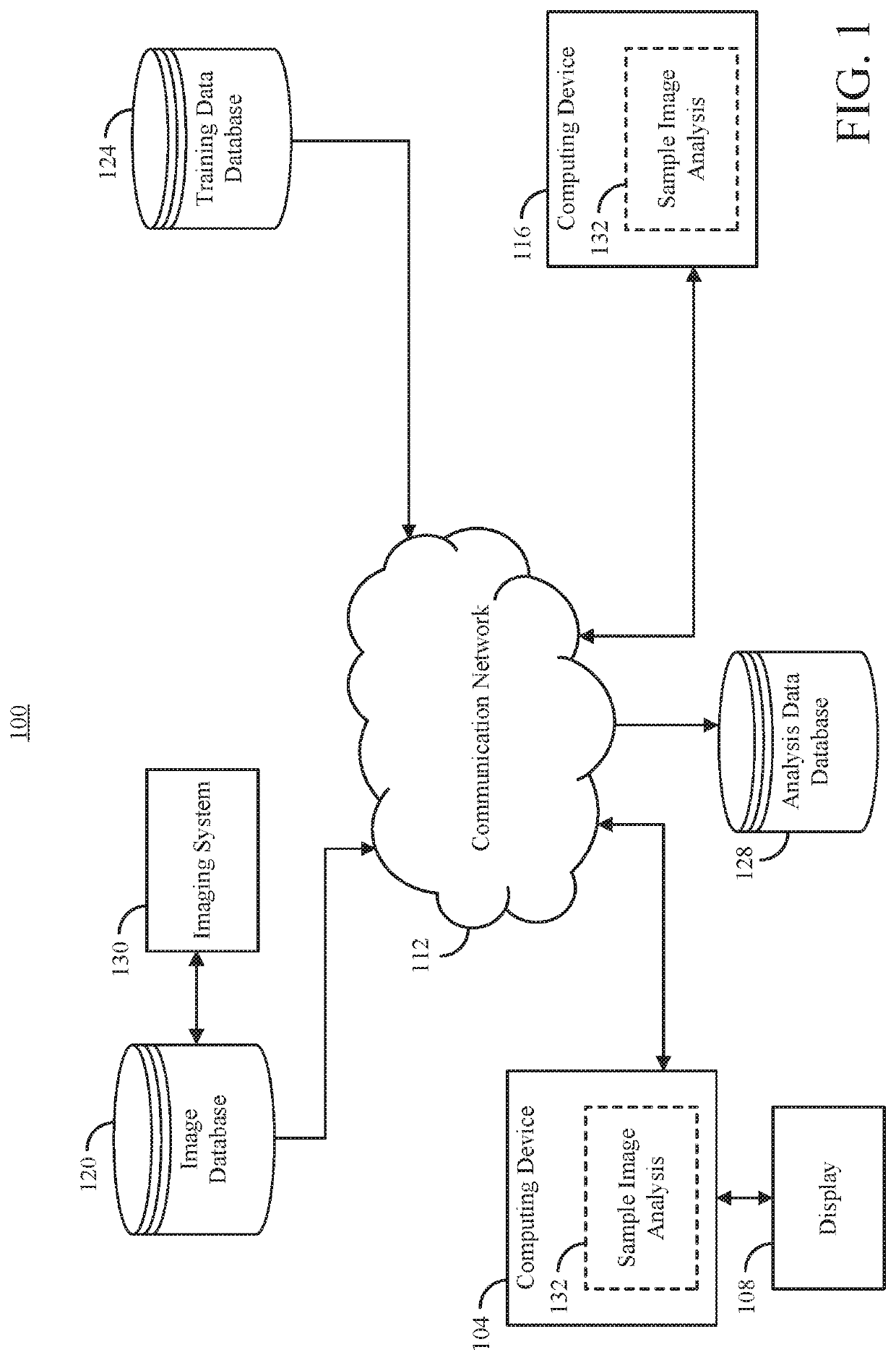
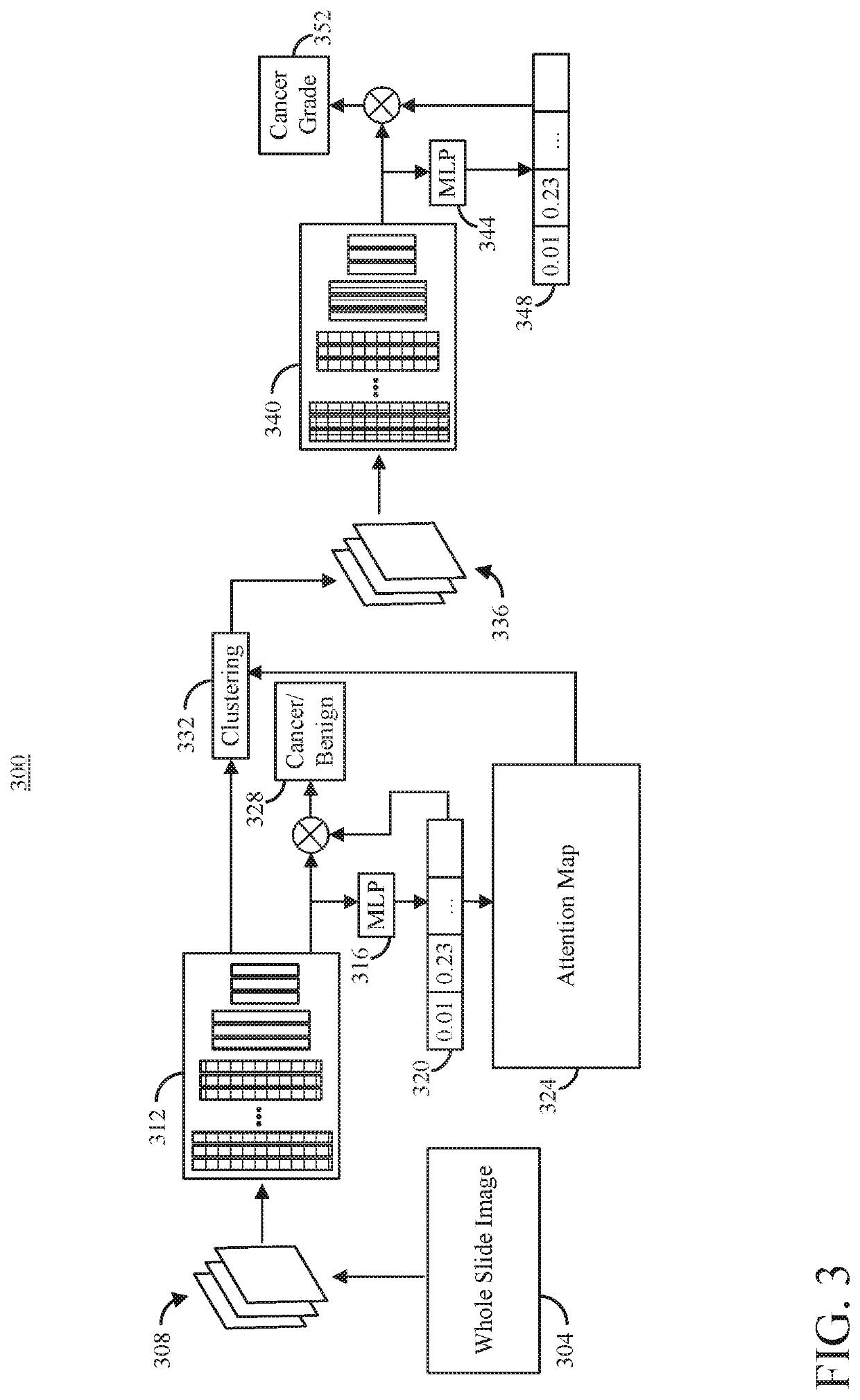
[0024]The present disclosure provides systems and methods that can reduce human and / or trained clinician time required to analyze medical images. As one non-limiting example, the present disclosure provides example of the inventive concepts provided herein applied to the analysis of images such as brightfield images, however, other imaging modalities beyond brightfield imaging and applications within each modality are contemplated, such as fluorescent imaging, fluorescence in situ hybridization (FISH) imaging, and the like. In the non-limiting example of brightfield images, the systems and methods provided herein can determine a grade of cancer and / or cancerous regions in a whole slide image (e.g., a digital image of a biopsy slide).

[0025]In some configurations of the present disclosure, an attention-based multiple instance learning (MIL) model is provided that can predict slide-level labels, but also provide visualization of relevant regions using inherent attention maps. Unlike pr...


PUM


Abstract

In accordance with one aspect of the disclosure, an image analysis system is provided. The image analysis system includes at least one processor configured to access image tiles associated with a patient, each tile comprising a portion of a whole slide image, individually provide a first group of image tiles to a first trained model, receive a first set of feature objects from the first trained model, cluster feature objects from the first set of feature objects to form a number of clusters, calculate a number of attention scores based on the first set of feature objects, select a second group of tiles, individually provide the second group of image tiles to a second trained model, receive a second set of feature objects from the second trained model, generate a cancer grade indicator, and cause the cancer grade indicator to be output.

Description

CROSS-REFERENCE TO RELATED APPLICATIONS[0001]This application is based on, claims the benefit of, and claims priority to U.S. Provisional Application No. 62 / 852,625, filed May 24, 2019, which is hereby incorporated by reference herein in its entirety for all purposes.STATEMENT REGARDING FEDERALLY SPONSORED RESEARCH[0002]This invention was made with government support under Grant Number CA220352, awarded by the National Institutes of Health. The government has certain rights in the invention.BACKGROUND OF THE INVENTION[0003]Medical imaging is a key tool in the practice of modern clinical medicine. Imaging is used in an extremely broad array of clinical situations, from diagnosis to delivery of therapeutics to guiding surgical procedures. While medical imaging provides an invaluable resource, it also consumes extensive resources. Furthermore, imaging systems require extensive human interaction to setup and operate, and then to analyze the images and make clinical decisions.[0004]As ju...

Claims


Application Information

Patent Timeline
IPC IPC(8): G06T7/00G16H30/20G16H15/00
CPCG06T7/0012G16H30/20G06T2207/20081G06T2207/30024G06T2207/20084G16H15/00G16H30/40G16H50/30G06N3/082G06T2207/30096G06T2207/20021G06V10/454G06N3/045G06F18/24133G06F18/23213
Inventor ARNOLD, COREYLI, JIAYUNSPEIER, WILLIAMLI, WENYUN
Owner RGT UNIV OF CALIFORNIA
Why Eureka
Try Eureka