Methods and materials for targeted expansion of regulatory t cells
a technology of regulatory t cells and materials, applied in the direction of peptide/protein ingredients, drug compositions, immunological disorders, etc., can solve the problems of toxicities and unsatisfactory off-target effects
- Summary
- Abstract
- Description
- Claims
- Application Information
AI Technical Summary
Benefits of technology
Problems solved by technology
Method used
Image
Examples
example 1
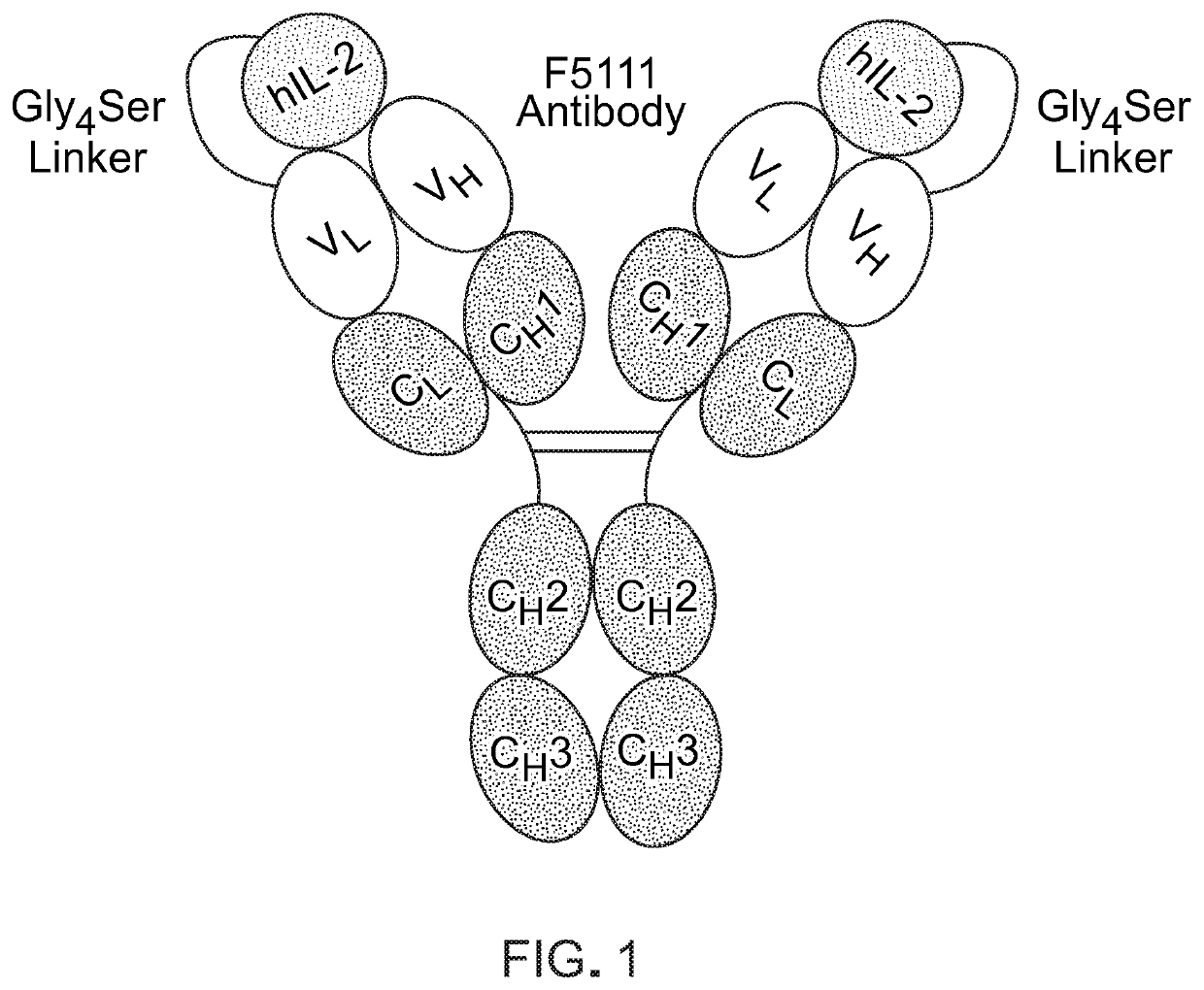
[0086]Engineered cytokine / antibody fusion for targeted expansion of human regulatory T cells
[0087]Administration of human IL-2 (hIL-2) in complex with the F5111 antibody was found to expand TRegS but not effector T cells from human peripheral blood and in humanized mouse models, presenting an enticing opportunity for targeted cytokine therapy. It was further shown that IL-2 / F5111 complex treatment reduces T1D severity in mice (Trotta et al., Nat Med. 24(7):1005-1014 (2018)). This exciting targeted IL-2 therapy holds vast clinical potential, but therapeutic development of a mixed cytokine / antibody complex is limited by dosing ratio considerations and complex instability. In fact, dissociation of the complex could induce dangerous toxicities from the free cytokine and potentially even exacerbate autoimmune pathogenesis by activating autoreactive effector T cells. Moreover, the free cytokine clears in J Immunol. 130(5):2203-2208 (1983)).
[0088]This Example describes the design and engin...
example 2
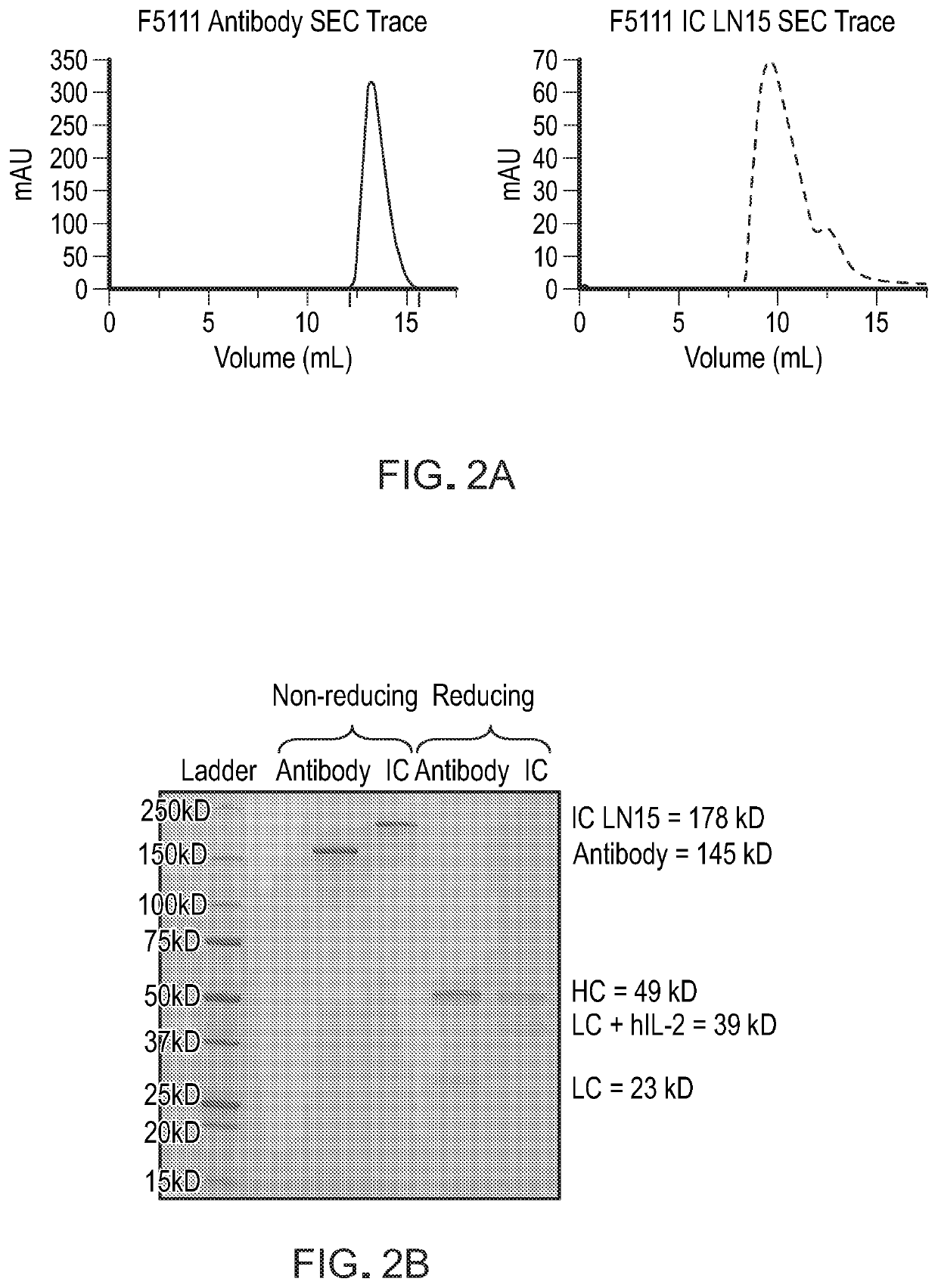
[0090]This example demonstrates that the recombinantly expressed single-chain F5111 IC is properly assembled and does not bind to IL-2Rβ.
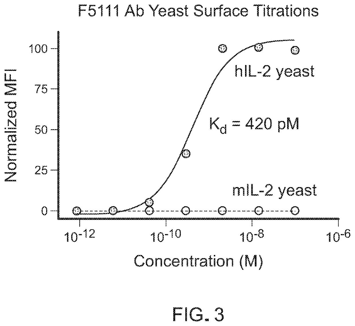
[0091]To demonstrate that hIL-2 is intramolecularly bound to the F5111 antibody within the IC, the binding affinity of F5111 IC LN15 for hIL-2 was measured and compared to that of recombinant F5111 antibody (Ab). The binding of purified F5111 antibody and IC to yeast surface-displayed hIL-2, as measured by flow cytometry, is shown in FIG. 4A. The binding affinities were also evaluated using bio-layer interferometry on an OCTET® instrument with biotinylated hIL-2 immobilized on a streptavidin-coated tip (FIG. 4B). For both yeast surface and bio-layer interferometry studies, a significant reduction in binding affinity was observed for F5111 IC LN15 relative to F5111 Ab, confirming intramolecular cytokine / antibody assembly. Bio-layer interferometry based binding studies were also conducted to assess the interaction between F5111 IC LN15 and the IL-2Rα...
example 3
[0092]This example demonstrates that the immunocytokine selectively biases IL-2 signaling.
[0093]IL-2 signals through activation of signal transducer and activator of transcription 5 (STAT5), which translocates to the nucleus to effect transcriptional programs (see, e.g., Murray, P.J. J Immunol, 178(5): 2623-2629 (2007); and Bromberg, J., and Wang, T.C., Cancer Cell, 15(2): 79-80 (2009)). To characterize IC variant-mediated immune bias, the YT-1 human NK cell line, which inducibly expresses the IL-2Rα subunit was employed (see, e.g., Yodoi et al., J Immunol, 134(3): 1623-1630 (1985)). Flow cytometry-based studies were performed to quantify STAT5 signaling elicited by IL-2, the IL-2 / F5111 complex, and F5111 IC LN15 on induced IL-2Rα+ versus uninduced IL-2Rα− YT-1 cells as a surrogate for TReg versus immune effector cell activation (FIG. 6). Untethered IL-2 signals potently on both IL-2Rαa+and IL-2Rα−cells, and IL-2 / F5111 complex fully activated both IL-2Rα+ cells and IL-2Rα− cells. In...
PUM
| Property | Measurement | Unit |
|---|---|---|
| Fraction | aaaaa | aaaaa |
| Fraction | aaaaa | aaaaa |
| Time | aaaaa | aaaaa |
Abstract
Description
Claims
Application Information
 - R&D
- Intellectual Property
- Life Sciences
- Materials
- Tech Scout
- Unparalleled Data Quality
- Higher Quality Content
- 60% Fewer Hallucinations
Browse by: Latest US Patents, China's latest patents, Technical Efficacy Thesaurus, Application Domain, Technology Topic, Popular Technical Reports.
© 2025 PatSnap. All rights reserved.Legal|Privacy policy|Modern Slavery Act Transparency Statement|Sitemap|About US| Contact US: help@patsnap.com


