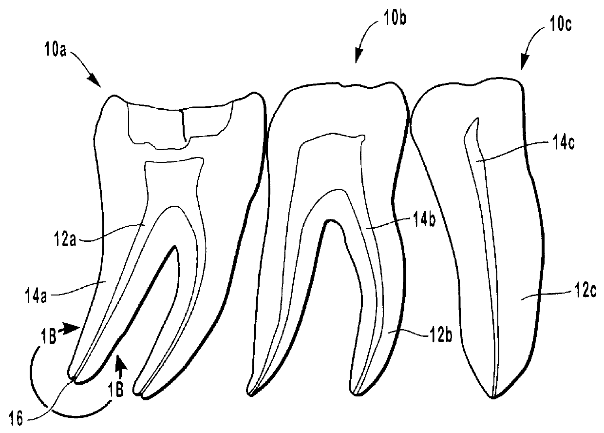
Although, x-
ray images obtained as shown in FIG. 2 from a buccal-lingual x-
ray projection are generally useful for determining the overall characteristics of a tooth, the approximate initial length of the root canal(s), and the working length for a file, such images provide only limited information regarding the overall
anatomy of the root canal.
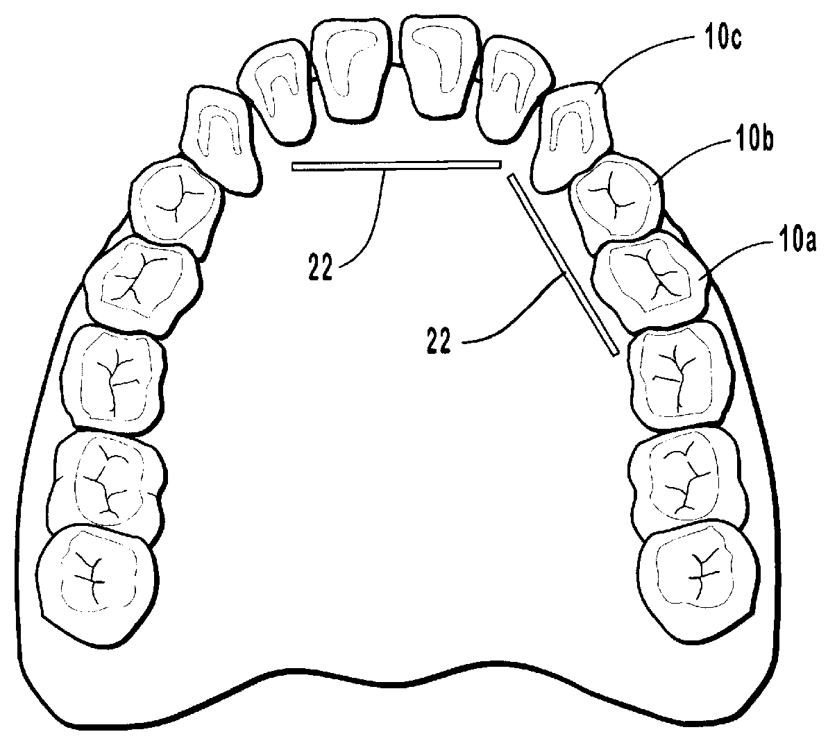
Although, it would be very helpful to view a tooth from a position between the teeth or from the
interproximal space such a mesial-distal view cannot be clearly produced when the tooth is still positioned in a patient's mouth.
More particularly, if not properly evaluated, x-
ray images can be misleading as to the
actual length of the root canal and the position of the
foramen or foramina.
However, we can never obtain a three-dimensional image!
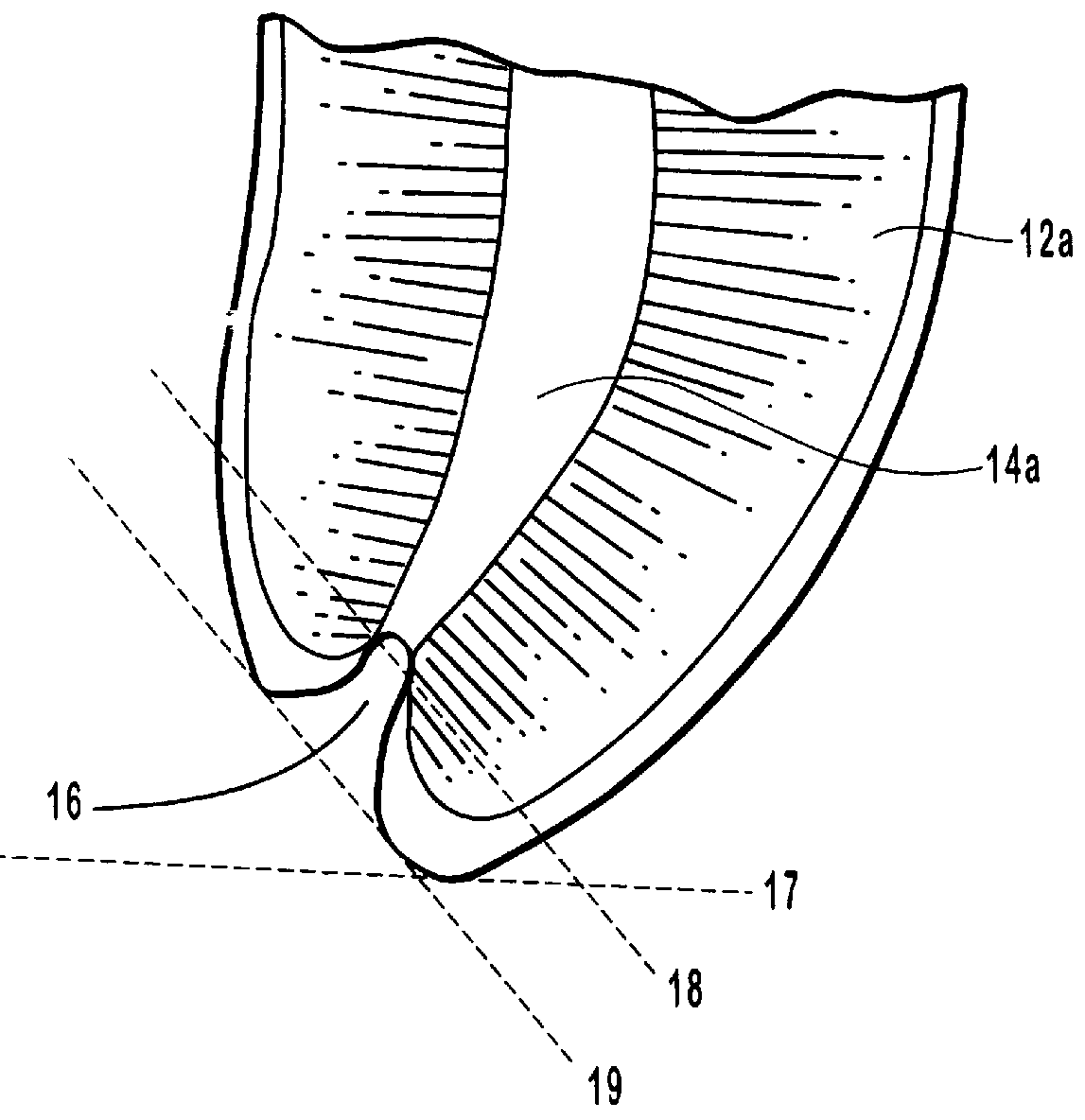
Comparing FIG. 3A with FIG. 3B clearly shows that when limited to knowledge derived from an x-ray corresponding to the image shown in FIG. 3A, the practitioner may not be able to accurately assess the anatomical structure of the root
anatomy.
More particularly, by comparing FIG. 3A with FIG. 3B or FIG. 4A with FIG. 4B, it is easily understood that the practitioner may not be able to accurately assess the anatomical structure of the root anatomy when limited to knowledge derived from an x-ray image.
Since it is impossible to obtain a mesial-distal view of the root canal or to view the perimetrical anatomy on different points along the length of the root canal, the practitioner is prevented from obtaining a proper preliminary understanding of the overall root canal anatomy in order to assess the necessary relationship between the canal walls and the instrument inserted in the root canal.
If the operator is unable to apply the instrument to every segment of the perimeter of the canal, the undisturbed pulp material may ultimately cause undue pain, lengthy healing times or even cause the procedure to fail.
In order to reach the recesses with filling material that cannot be treated with the instruments, vertical pressure must be applied with a plugger; however, there is never any assurance that all of the necrotic residue has been coated.
There is also a risk that such techniques will cause infected material to be pressed beyond the apex.
Such an
extrusion of infected material beyond the apex is very undesirable as it may contain polymicrobial loads or charges that may produce damaging
bacteremia or cause
chronic inflammations of the apical and periapical tissues.
Based on all of the foregoing observations, it can be concluded that inadequate attention is given to understanding the dental chambers on a three-dimensional basis, the varying configurations of the perimeter of the root canal(s), the
diameter of the canals, and the thickness of the walls.
The inability to fully identify the anatomy of the pulp cavity restricts the ability of the practitioner to confidently conclude that the procedure has been successful.
Although problems may result from having incomplete information regarding the anatomy of a particular root canal, many practitioners using conventional methods and instruments are not overly concerned with completely cleaning the entire root canal since their
failure rate is not at an unsatisfactory level.
As a result, it becomes increasingly difficult for the files to adjust to or to follow the contours of the perimeter surfaces of the root canal.
This reduced flexibility also increases the likelihood that the files will fail to contact some portions while removing too much of the surrounding
dentin 154 in some areas through excessive abrasion and resulting in overthinning of the walls.
There is resultingly some possibility for failure of the root canal therapy due to incompleteness.
Not only is the completeness effected by the use of a set of files wherein each file is more rigid than the preceding file but the ability to safely move the file within the canal is also limited.
More particularly, the increasing rigidity results in decreased ability to negotiate the curves in the canal.
Significant problems that can result from inserting increasingly rigid files and also from initially inserting a file all the way down to the apex includes laceration and transportation of the apical
foramen, as well as misdirection and perforation of the wall.
Perforating the apex can also result from an error in estimating the length of a root canal, by failure of a stop such as stop 140 to remain at a predetermined position or by failure to observe the calibration or graduation hatch markings on the file, which can be used instead of a stop to designate the length.
As a result, the periapical region can be invaded and contaminated.
The potential for extruding infected material through the apical
foramen of a necrotic tooth during the initial
insertion of a file instrument all the way down to the apex is a particular
disadvantage of the step-back technique.
Another
disadvantage is that the procedure has identical steps for working in either necrotic or vital root canals.
Such apical perforations, as well as wall perforations, may
delay tooth healing and may compromise the outcome of the therapy.
Perforations can also occur due to a failure to maintain a proper working length of the instrument during the procedure.
The time required to obtain the x-ray photographs or images and to adjust the working length of the instruments by repositioning the stops can result in a lengthy process.
The step-back technique is also time intensive because a large number of instruments are required to complete the root canal therapy.
Such ledges are difficult to bypass; and if the ledge occurs very close to the apex, the ledge may give the practitioner the mistaken impression that the apex has been reached.
However, as discussed hereinbelow, significant potential problems may inherently result from use of the crown-down technique.
One example of the operational deficiency of the crown-down method lies in its association with instruments made of
nickel /
titanium.
Additionally, it has been asserted that such files are more likely to stay in the center of the root canal, thereby decreasing the likelihood of ledging or perforating the root canal walls.
The ability of a
nickel /
titanium file to stay in the center is not necessarily desirable, in view of the morphology and perimetrical variety of root canals, and particularly the variety in the upper two-thirds of laminar root canals.
Moreover, because
nickel /
titanium files are more flexible than steel files, they tend to follow the path of least resistance and therefore cannot be used, in the same way as steel files, to be applied actively and intentionally by the operator.
As a result, even when the operator knows the thickness of a particular portion, such as an interference or obstruction which the operator desires to rectify or straighten, the operator lacks the freedom to aggressively drive the file as needed and clean the portions that are difficult to reach.
Accordingly, when a nickel / titanium file is used to clean a non-cylindrically shaped root canal, the file moves only at the center of the canal and / or the area of least resistance and fails to remove all of the
necrotic tissue.
These problems can be easily caused by the passive, self-guiding use of nickel / titanium files with progressively larger tapers in the transition from the first instrument to the next one in the set, and increasing rigidity in accordance with the crown-down technique which prevents the files from being laterally moved to enable the files to clean the entire perimeter of the root canal.
Such overthinning and potential furcal perforation can have devastating results.
The inability to adequately direct a file used in accordance with the crown-down technique based on the practitioner's knowledge of the relative thicknesses of the portions of canal walls is a significant
disadvantage of the technique.
As a result, the practitioner may not realize that the borehole extends into the
cementum and may therefore mistakenly conclude that the root canal treatment has been successful.
Infective
bacteria that remained in the root canal, perhaps in the portions that were not contacted with the files, as well as toxins produced by the
bacteria may then permeate through the
cementum and cause infection or other complications.
Since the practitioner is prevented from removing and cleaning essentially all pulp material, the practitioner cannot be assured of the reliability of the treatment.
Additionally, the practitioner may not suspect that the working instruments have failed to contact every segment of the root canal as use of a set of files with increasingly greater tapers can contribute to a potentially incorrect conclusion that cleaning by such a conventional process has resulted in removing all material from root canal 194a.
It should also be remembered that while rotation of a set of passively actuated files, with increasingly greater tapers, in the center of the canal, in accordance with the crown-down technique, may yield a configuration as shown in FIG. 15D and result in successful root canal therapy, there is a significant
hazard, as shown in relation to the FIGS. 15A-C, due to the passivity of the instruments when linked to canal diameters and wall thicknesses that are still statistically unknown.
The excessive
thinning of the dental wall may resultingly significantly weaken the resistance of the walls to the stress of chewing, and may also cause a fracture of the root.
From the above discussion in relation to FIGS. 15A-E, it is clear that the actual morphology of the canals is not sufficiently considered when using this method and that the use of files with increasingly larger tapers limits the
range of motion of the files.
More specifically, due to the use of files with successively larger tapers which therefore are increasingly rigid, each file, if actuated passively, is primarily limited to being rotated without substantial lateral movements guided by the operator.
Since the majority of files are of the laminar type, this limitation poses a significant problem.
Without the ability to laterally move the files, it is not possible to make contact with every segment of the perimeter of the canal and some portions may receive too much contact.
Accordingly, the files cannot clean a root canal without significantly altering the original anatomy by leaving a
footprint or borehole corresponding to the configuration of the instruments used.
More specifically, the result is a
footprint or borehole with a perimeter that corresponds to the perimeter of the biggest file that extends well beyond the original anatomy of the root canal and yet in most instances does not adequately clean significant portions of the root canal.
There are also other disadvantages to the use of nickel / titanium files.
Since nickel / titanium files are more fragile and more flexible than stainless steel files, the nickel / titanium files can break more easily and unexpectedly than steel files.
More particularly, beyond a certain
diameter, the upper halves of larger
diameter files are still as rigid as that of steel files while the flexible lower halves of nickel / titanium file instruments are more prone to break.
Additionally, rotation of a file in a canal that has a laminar upper two-thirds exposes the tip of the file to the risk of breaking when the tip of the file is embedded or stuck in a canal whose diameter is smaller than its own diameter!
Another disadvantage of nickel / titanium files is that nickel embodied in the
alloy may potentially result in an
allergic reaction.
Further, nickel / titanium files costs about four times as much as steel files and yet nickel / titanium files generally
wear out faster than steel files.
Nickel / titanium files
wear out so quickly that some manufacturers mark their products as being intended for
single use only.
Moreover, the crown-down instruments currently available on the market, almost all of which are made of nickel / titanium, in some respects violently conflict with the use of the crown-down method, because, paradoxically, these instruments are smooth in areas where the method requires that they first perform a
cutting action.
The need to frequently change the cleaning instrument results in significant time requirements for cleaning a root canal.
However, careful
instrumentation in accordance with either tedious
time consuming method does not avoid the problems set forth above in relation to apical perforation, wall perforation, overthinning or failure to clean all of the wall surfaces.

