System and method for fusing three-dimensional shape data on distorted images without correcting for distortion

a three-dimensional shape and image technology, applied in the field of robotic and medical imaging techniques, can solve the problems of inconvenient and costly patient removal of pins, no conventional technique teaches how to simulate post-operative conditions, and the insertion of pins requires minor surgery

Inactive Publication Date: 2002-07-02
IBM CORP
View PDF10 Cites 104 Cited by
  • Summary
  • Abstract
  • Description
  • Claims
  • Application Information

AI Technical Summary

Benefits of technology

Another object of the present invention is to provide a system and method for fusing three-dimensional shape data on distorted images without correcting for distortion.
Further, with the invention, a clinician or surgeon can view images in the manner that they are accustomed. That is, the clinician or surgeon can view the unmodified image (e.g., an image with distortion), in the manner with which they are familiar. Thus, the surgeons can continue to interpret the unmodified images, as is customary. Further, since no correction is performed as in the conventional system and methods, no image degradation or blurring results from such image correction and reformatting of the image. Additionally, processing speed is not decreased, and similarly processing resources are not increased since the processing of the method of the present invention is less complex than that of the conventional systems and methods.

Problems solved by technology

However, the insertion of the pins requires minor surgery.
However, using such pins as markers is not always desirable, as they may cause significant patient discomfort, and the required surgical procedure to insert and subsequently remove the pins is inconvenient and costly to the patient.
However, none of the conventional techniques teaches how to simulate a post-operative condition depending upon the registration of image data to physical space, by fusing intra-operative images with registered pre-operative data, and generating new images.
Herring et al. do not teach how to evaluate the registration accuracy intra-operatively.
With Taylor's system, a surgeon can take corrective measures to minimize the effects of a wrongful execution of the plan, but cannot make a decision before any execution of the plan and therefore cannot prevent all errors before they occur.
Further, the information produced by Taylor's system for advising a surgeon is not represented in the form of conventional medical media (e.g., such as X-ray images) and requires an extra burden on the surgeon in order to interpret and evaluate this information.
Thus, it is believed that conventional techniques do not exist (or at the very least are inadequate) for (a) providing the surgeon with post-operative evaluations prior to surgery, that are obtained by merging intra-operative image data and pre-operative data, and (b) presenting such evaluations in a standard clinical fashion (e.g., such as augmented X-ray images) that is natural for a surgeon to interpret.
Other problems of the conventional systems and methods include the limited availability of 2-D / 3-D registration methods in conventional art systems for advising a surgeon.
This is problematic because a clinician or surgeon is used to seeing the unmodified image (e.g., an image with distortion).
That is, as a practical matter, surgeons generally are not familiar with seeing the modified image.
Further, such a correction may cause image degradation or blurring due to the reformatting of the image.
Additionally, slower and more complex processing results from the image correction process.

Method used


Image

  • System and method for fusing three-dimensional shape data on distorted images without correcting for distortion

Examples


Embodiment Construction

Referring now to the drawings, and more particularly to FIGS. 1-8, there is shown a preferred embodiment of the method and structure according to the present invention.

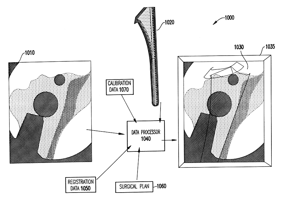
Generally, the present invention resides in a system and method to intra-operatively provide the surgeon with visual evaluations of possible surgical outcomes ahead of time, the evaluations being obtained by merging intra-operative image data and pre-operative data, and being presented in a standard clinical fashion (such as augmented X-ray images) that is natural and easy for a surgeon to interpret.

The present invention differs from the invention in U.S. patent application Ser. No. 09 / 299,643 by omitting the step of correcting the geometric distortion of the X-ray image in the method of U.S. patent application Ser. No. 09 / 299,643 (e.g., step 2040 in FIG. 2 thereof) and other processing as described below.

That is, the step of correcting the geometric distortion of the X-ray image is omitted between the step of obtaini...


PUM


Abstract

A system and method for intra-operatively providing a surgeon with visual evaluations of possible surgical outcomes ahead of time, and generating simulated data, includes a medical imaging camera, a registration device for registering data to a physical space, and to the medical imaging camera, and a fusion mechanism for fusing the data and the images to generate simulated data, without correcting for distortion. The simulated data (e.g., such as augmented X-ray images) is natural and easy for a surgeon to interpret. In an exemplary implementation, the system preferably includes a data processor which receives a three-dimensional surgical plan or three-dimensional plan of therapy delivery, one or a plurality of two-dimensional intra-operative images, a three-dimensional model of pre-operative data, registration data, and image calibration data. The data processor produces one or a plurality of simulated post-operative images, without correcting for distortion, by integrating a projection of a three-dimensional model of pre-operative data onto one or a plurality of two-dimensional intra-operative images.

Description

1. Field of the InventionThe present invention generally relates to robotics and medical imaging techniques and, more particularly to robotically-assisted surgical systems and other devices incorporating methods for registering image data (both pre-operative and intra-operative) to physical space and for providing feedback, and in particular visual feedback, to the clinician.2. Description of the Related ArtComputers are increasingly used to plan complex surgeries by analyzing pre-operative Computed Tomography (CT) or Magnetic Resonance Imaging (MRI) scans of a patient.To execute the surgical plan, it is important to accurately align or register the three-dimensional pre-operative and intra-operative data to an actual location of the patient's anatomical features of interest during surgery.One conventional technique for performing this type of registration is to attach a stereo-tactic frame or fiducial markers to the patient, and to precisely locate the frame or markers prior to and...

Claims


Application Information

Patent Timeline
Patent Type & Authority Patents(United States)
IPC IPC(8): A61B6/03
CPCA61B6/032A61B6/463
Inventor GUEZIEC, ANDREKALVIN, ALAN DAVIDWILLIAMSON, JR., WILLIE
Owner IBM CORP
Why Eureka
Try Eureka