
Epitopes in viral envelope proteins and specific antibodies directed against these epitopes: use for detection of HCV viral antigen in host tissue

a technology of hcv and specific antibodies, which is applied in the field of hcv infection and hcv virus antigen detection, can solve the problems of false positive reactivity, cumbersome rna detection in cells or tissues, and major health problems

Inactive Publication Date: 2003-02-18
INNOGENETICS NV (NL)
View PDF16 Cites 9 Cited by
  • Summary
  • Abstract
  • Description
  • Claims
  • Application Information

AI Technical Summary

Benefits of technology

It is clear from the literature that there is an urgent need to develop reliable diagnostic methods, reliable vaccines and effective therapeutic agents for HCV. Also sensitive and specific screening methods of HCV-contaminated blood-products and improved methods to culture HCV are needed. New antibodies able to detect the virus in animal--or in vitro models, or in its natural host, may help in designing efficient diagnostic tools and therapeutic agents. In this regard, the present invention is based on the surprising finding of monoclonal antibodies directed against either E1 or E2-HVR which can be used for the detection of HCV antigens in various ti

Problems solved by technology

Hepatitis C virus (HCV) infection is a major health problem in both developed and developing countries.
However, RNA detection in cells or tissues is a cumbersome technique which either involves the extraction of RNA followed by reverse transcription and nested PCR or includes in situ RT-PCR and hybridisation.
Since these techniques are also prone to false positive reactivity, viral RNA detection is exclusively performed on serum samples.
Reliable methods for the detection of viral protein antigens in serum and tissue samples are still lacking.
The detection of viral proteins in cells has been hampered by the lack of antibodies which specifically bind to viral proteins and which are able to specifically recognise the natural HCV protein antigens (i.e. antigens as expressed by the host following infection with HCV).
Preparations containing host-derived antibodies, however, cannot be reproduced easily and these preparations may be contaminated by autoimmune antibodies as well as by antibodies against other known or even unknown agents.
Currently, this expression condition goes together with incomprehensible problems.
Thus, routine detection of HCV antigen in liver biopsies still lacks sensitivity.
Also the detection of viral antigens in body fluids such as serum or plasma is hampered by the same problem.
Thus, identification of such epitopes and the search for antibodies with high cross-reactivity towards the sequence variation of HCV is a challenging undertaking.

Method used


Image

  • Epitopes in viral envelope proteins and specific antibodies directed against these epitopes: use for detection of HCV viral antigen in host tissue
  • Epitopes in viral envelope proteins and specific antibodies directed against these epitopes: use for detection of HCV viral antigen in host tissue
  • Epitopes in viral envelope proteins and specific antibodies directed against these epitopes: use for detection of HCV viral antigen in host tissue

Examples


example 1

Generation of Monoclonal Antibodies Against E1 and E2

Mice were immunised with truncated versions of E1 (aa 192-326) and E2 (aa 384-673), expressed by, recombinant vraccinia virus as described in PCT / EP 95 / 03031 to Maertens et al. After immunisation splenocytes of the mice were fused with a myeloma cell line. Resulting hybridomas secreting specific antibodies for E1 or E2 were selected by means of ELISA.

example 2

Selection of Monoclonal Antibodies

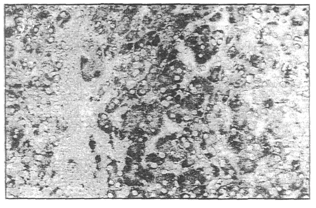
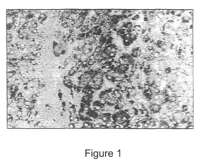
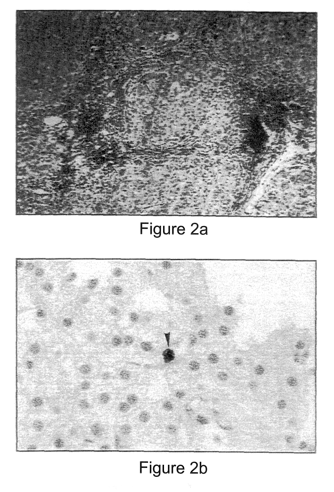
A large series (25 in total, of which 14 in detail; the latter can be obtained from Innogenetics NV, Gent, Belgium) of monoclonals directed against E1 or E2 was evaluated for staining of native HCV antigen in liver biopsies of HCV patients and controls (see below). Staining was performed either on cryosections or on formadehyde fixed biopsies (for protocols. see figure legends). Only three monoclonal antibodies revealed a clear and specific staining. All other monoclonal antibodies either gave no or very weak staining, or showed non-specific staining,. Remarkably, two different antigen staining patterns were noticed. Monoclonal IGH 222, directed against E2, clearly stained hepatocytes (FIG. 1), while IGH 207 and IGH 210, directed against E1, stained lymphocytes infiltrating in the liver (FIGS. 2a and 2b). IGH 207 also stained hepatocytes, but to a weaker degree than IGH 222 (compare FIGS. 1 and 2a). This-staining pattern was confirmed on a series of...

example 3

Identification of Monoclonal Antibodies Allowing Detection of Viral Envelope Antigen in Peripheral Blood Cells

The finding that lymphocyte infiltrates in the liver can be stained for HCV envelope antigens prompted us to look also at peripheral blood mononuclear cells. In order to allow intracellular staining, peripheral blood cells were permeabilised with saponin, allowed thereafter to react with the monoclonal antibodies IGH 201 or 207 (directed against E1 and showing weak and strong positivity on the liver biopsies, respectively). Finally, reactivity was checked on a fluorescent cell sorter using secondary FITC-labelled antibodies. IGH 207, which stained already the lymphocyte infiltrates in the liver, showed a high specificity. With this monoclonal, almost no background staining was detected, and 4 out of 5 patients clearly stained positive (FIG. 3). The second monoclonal, IGH 201 (binding to SEQ ID 29; ECACC accession number: 98031216) which is also directed against E1, yields a ...


PUM


Abstract

Antibodies to two new epitopes on the HCV envelope proteins were identified which allow routine detection of native HCV envelope antigens, in tissue or cells derived from the host. The new epitopes are: the E1 region aa 307-326 and the N-terminal hyper variable region of E2 aa 395-415. Surprisingly, we characterised an antibody that reacts with various sequences of the hypervariable domain of E2. Specific monoclonal antibodies directed against these epitopes and allowing routine detection of viral antigen are described.

Description

The present invention is based on the finding that antibodies directed against specific epitopes of the E1 and E2 protein of HCV can be used to detect viral antigens in host tissues.Hepatitis C virus (HCV) infection is a major health problem in both developed and developing countries. It is estimated that about 1 to 5% of the world population is affected by the virus. HCV infection appears to be the most important cause of transfusion-associated hepatitis and frequently progresses to chronic liver damage. Moreover, there is evidence implicating HCV in induction of hepatocellular carcinoma. Consequently, the demand for reliable diagnostic methods and effective therapeutic agents is high. Also sensitive and specific screening methods of HCV-contaminated blood-products and improved methods to culture HCV are needed.HCV is a positive stranded RNA virus of approximately 9,400 bases which encode at least three structural and six non-structural proteins. Based on sequence homology, the str...

Claims


Application Information

Patent Timeline
IPC IPC(8): C07K14/18C07K14/005C07K16/08C07K16/10C12N5/10C12N5/20C12N15/02G01N33/576C12P21/08C12R1/91
CPCC07K14/005C07K16/109C12N2770/24222A61P31/14C07K16/10
Inventor MAERTENS, GEERTDEPLA, ERIKBUYSE, MARIE-ANGE
Owner INNOGENETICS NV (NL)
Learn More