Eureka AIR delivers breakthrough ideas for toughest innovation challenges, trusted by R&D personnel around the world.

Chest brace and method of using same

a brace and chin strap technology, applied in the field of chin straps, can solve the problems of weakened respiratory system of newborn infants, cpap, and weakened tidal volume, and achieve the effects of reducing tidal volume, serious side effects, and major limitations

Inactive Publication Date: 2003-03-18
PENN STATE RES FOUND +1
View PDF55 Cites 42 Cited by
  • Summary
  • Abstract
  • Description
  • Claims
  • Application Information

AI Technical Summary

Benefits of technology

It is another object of this invention to provide a chest brace that provides a continuous distending pressure on the patient's chest so as to correct any collapse that is evident between breaths, i.e., at the end of expiration.
It is still another object of this invention, to provide a chest brace that provides continuous distending pressure on the patient's chest cavity without requiring vacuum seals.
It is still another object of this invention to provide a chest brace that is simple to attach, inexpensive, and does not require electrical power to operate.
It is a further object of the present invention to provide a chest brace and method of using such a brace to improve a patient's thoracic index even in the absence of chest collapse associated with respiration.
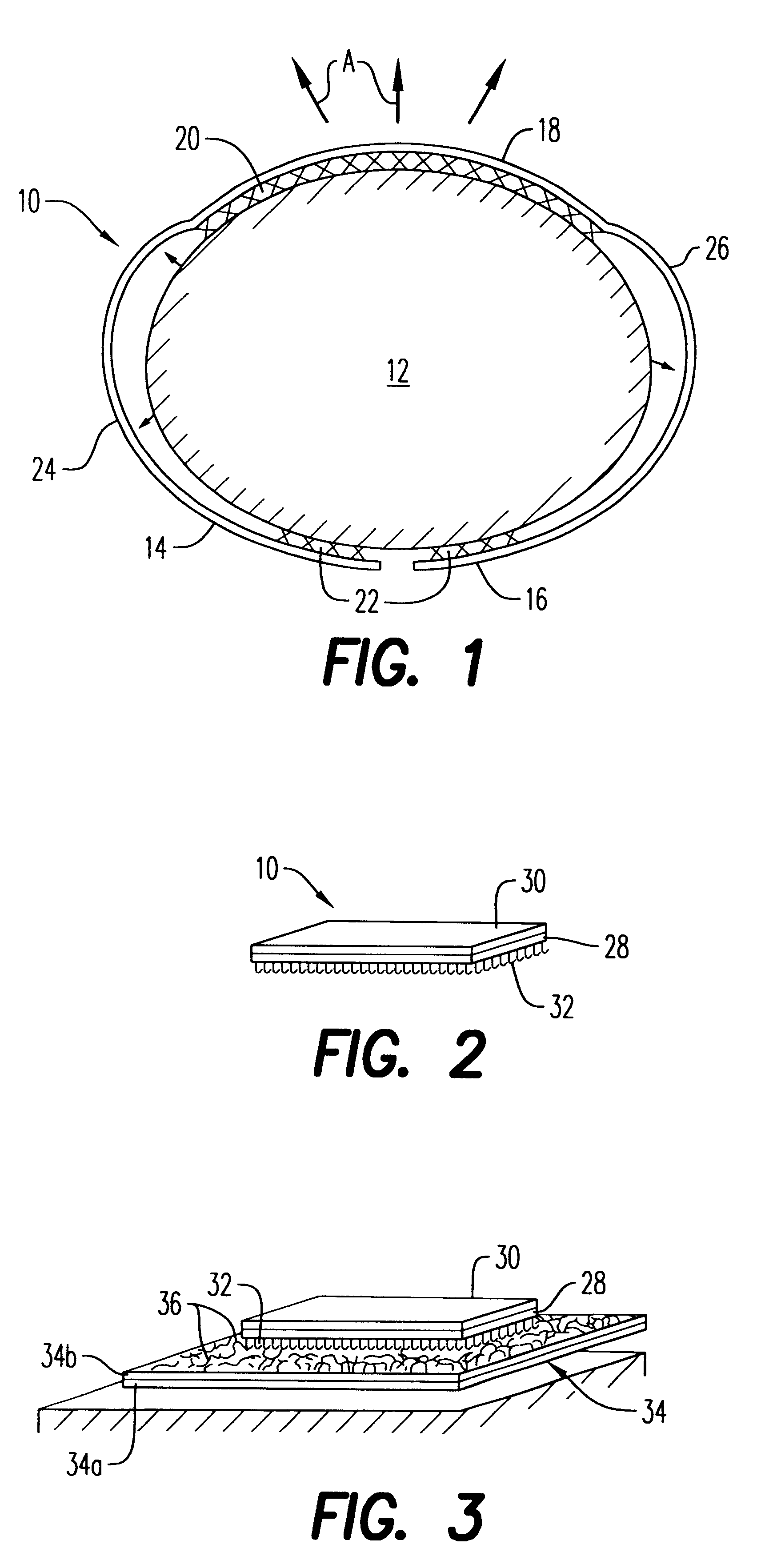
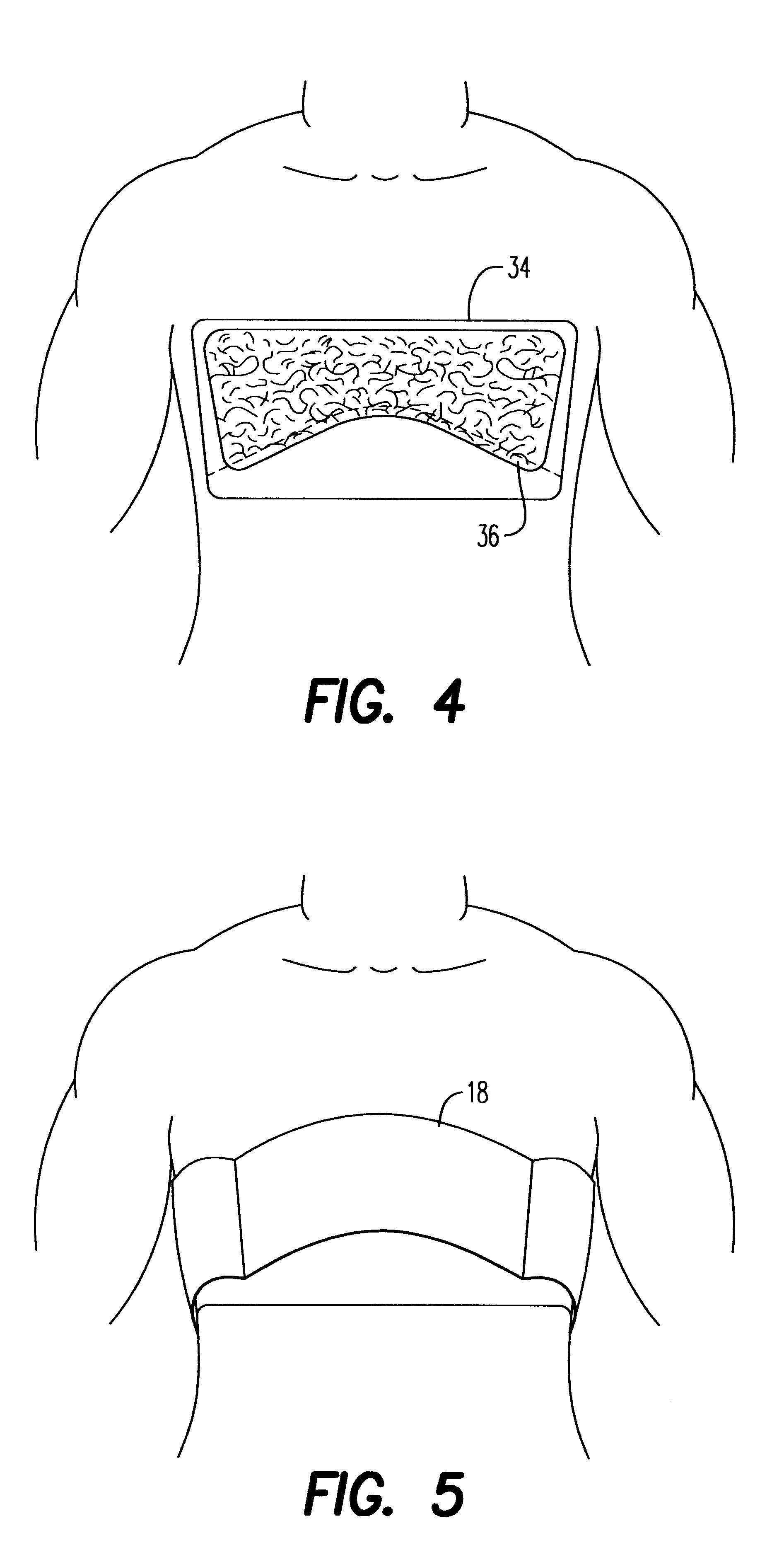
These objects, among others, are achieved, according to one embodiment of the present invention, by providing a chest brace that includes an anterior member adapted to overly a patient's chest. An adhesive mechanism secures the anterior member to the surface of the patient. A support structure is coupled to the anterior member such that, in use, the support structure imparts a force on the anterior member in a manner so as to distend a thorax of the patient. In one embodiment of the present invention, the support structure includes a posterior member that is coupled to the anterior member using a mechanical linkage, so that the anterior member can be moved relative to the posterior member in a ratchet-like fashion, thereby imparting the distending pressure. This allows the patient to receive the distending force regardless of whether they are in the supine or prone position. In another embodiment of the present invention, the support structure includes a support structure, such as a shield, having a portion that is placed over the patient. The anterior member is suspended from the shield in a manner so as to impart the distending force on the patient's chest. The present invention also contemplates adhering a chest plate to the patient so that the anterior member can be selectively attached to the patient merely by securing it to the chest plate, thereby avoiding the need to remove the adhesive from the patient each time the patient is removed from the chest plate.
It is a further object of the present invention to provide a method of imparting a distending force on a thorax of a patient that that overcomes the shortcomings of conventional methods for preventing collapse of a patient's chest, improving the patient's thoracic index, or both. This object is achieved by providing a method that includes adhesively securing an anterior member of a chest brace structure to a patient's chest and imparting a force on the anterior member so as to distend a thorax of such a patient. Numerous techniques for imparting a force on the anterior member are discussed in detail below.

Problems solved by technology

Pulmonary insufficiency associated with immaturity is one of the most common life-threatening hurdles that confronts the premature newborn baby.
Also, the relatively compliant chest wall tends to collapse as the diaphragm descends, resulting in a diminished tidal volume.
CPAP, however, is potentially hazardous to newborn infants with weakened respiratory systems.
It is usually administered by nasal prongs, but has major limitations and serious side effects.
These include: nasal trauma, difficulty in obtaining a good fit in very small infants, and high gas flows that cause airway cooling, drying, and obstruction of the nasal passages.
During periods of crying and mouth opening, especially with high CPAP flows, there is a loss of pressure and the infant inhales room air.
Frequent dislodgement of the nasal prongs makes nursing difficult, especially when associated with repeated bouts of desaturation.
High or fluctuating saturation may increase the risk of retinopathy.
Perhaps more serious are the circulatory disturbances, decreased venous return to the heart, diminished cardiac output, and increased intra-cranial hemorrhage.
While improvements have been made in the design of devices for generating extra-thoracic negative pressure, the devices are still difficult to attach to small newborns.
These devices require some form of electrical power supply, are relatively expensive, and are cumbersome.
Technical difficulties are associated with temperature control, neck seals obstructing venous return, leaks around the seals and limited patient access.
These devices also require considerable training and experience to operate and the technical problems make nursing difficult and frustrating.
This limits the use of a potentially life saving treatment modality.
However, conventional assisted ventilation and CNP devices, which used primarily to prevent lung collapse, does little or nothing to improve the infant's thoracic index.

Method used


Image

  • Chest brace and method of using same
  • Chest brace and method of using same
  • Chest brace and method of using same

Examples


second embodiment

In addition to providing rigidity for the patient's chest wall and a continuous negative distending pressure, a chest brace 10' is adapted to provide active ventilation. Referring to FIG. 8, the exterior surface of chest brace 10' includes an air bladder 50 that is bonded thereto. By controlling the amount of air within air bladder 50, via tube 52, the stiffness of bladder 50 can be altered to control the amount of outward pull of chest brace 10', as indicated by arrows B. More specifically, filling bladder 50 with air changes its shape, and as bladder 50 straightens, it pulls the brace away from the chest. When pressure is released from air bladder 50, chest brace 10' is enabled to resume its original position by the natural resiliency of its metal core. In such manner, ventilation of the patient can be assisted by periodically altering the air pressure within air bladder 50.

third embodiment

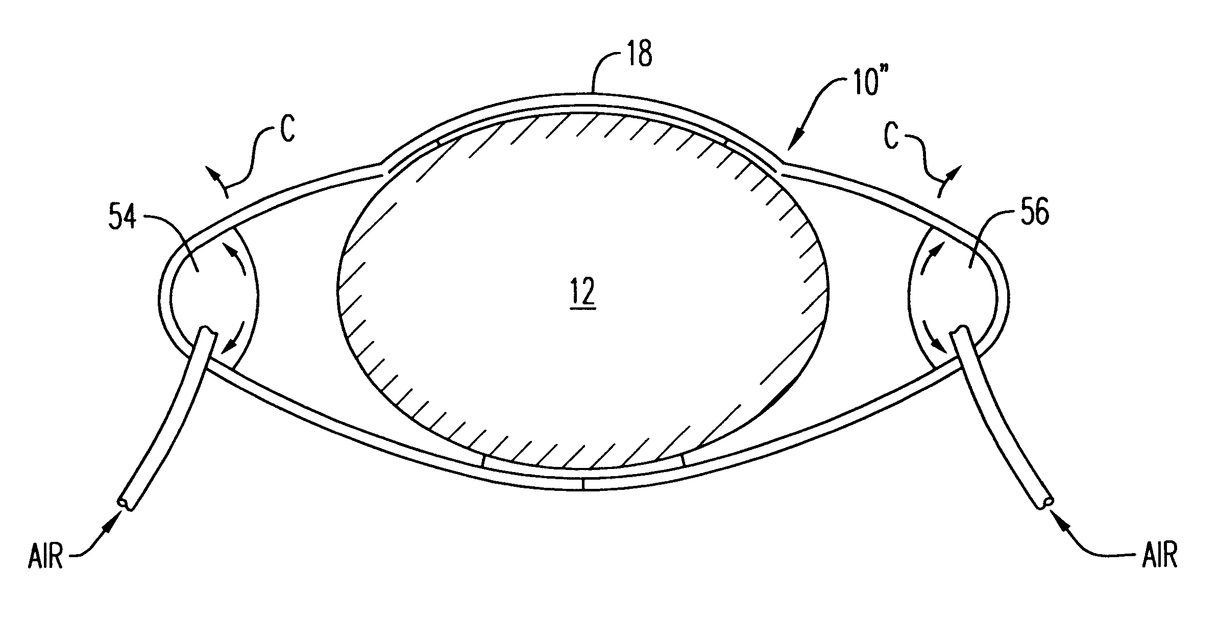
In FIG. 9, a chest brace 10" is shown. Chest brace 10" provides the same ventilation function as chest brace 10' of FIG. 8 except that in this case, a pair of bladders 54 and 56 are positioned within a chest brace 10". Inflation and deflation of the bladders, either individually or simultaneously, controls the position of frontal resilient segment 18 of chest brace 10", as generally indicated by arrows C. In such a manner, ventilation of the patient is assisted.

fourth embodiment

FIG. 10 illustrates a chest brace 60 that comprises a pair of separated brace members 62 and 64. Anterior brace member 62 is adhered to the patient's chest wall via the same connection mechanism as described above. Similarly, posterior brace member 64 is adhered to the back of the patient in the manner described above. The spacing between brace members 62 and 64 is controlled by air pressure within a pair of corrugated respirator tubes 65 and 66. Thus, as pressure is increased within corrugated tubes 65 and 66, anterior brace member 62 moves away from posterior brace member 64. Through the action of the Velcro interconnection between anterior brace member 62 and the patient's chest wall, the patient's chest wall moves outwardly. When, however, pressure is reduced within corrugated tubing 65 and 66, a vacuum is created thereby causing a squeezing action on the patient's chest between brace numbers 62 and 64. In such manner, the patient's respiration is assisted. Control of air pressu...


PUM


Abstract

A chest brace and method of using same that prevents the chest wall from buckling inwards during patient breathing by providing a distending force on the patient's thorax. The chest brace also restores the normal anterior-to-posterior chest dimensions by providing a distending force in this direction. The chest brace includes an anterior member adapted to overly a patient's chest. An adhesive mechanism secures the anterior member to a surface of such a patient. A support structure is coupled to the anterior member such that, in use, the support structure imparts a force on the anterior member in a manner so as to distend a thorax of such a patient.

Description

1. Field of the InventionThe present invention relates to a chest brace and method of using a chest brace to prevent collapse of a chest wall of a patient, such as a neonate, to keep the lungs inflated. More particularly, the present invention pertains to an inexpensive chest brace that interacts with the skin covering the chest, rather than through applied negative air pressure, to provide a distending force on the chest wall to prevent its collapse, especially during respiration.2. Description of the Related ArtPulmonary insufficiency associated with immaturity is one of the most common life-threatening hurdles that confronts the premature newborn baby. The newborn's rib cage is soft and buckles easily during spontaneous respiration, particularly during inspiration. Underdevelopment of the intercostal muscles, lungs, or both contributes to the chest's deformability. In premature infants below 30 weeks gestation, thoracic wall elastic recoil is almost non-existent, so that the rest...

Claims


Application Information

Patent Timeline
IPC IPC(8): A61H31/00A61F5/00
CPCA61F5/03A61H31/00A61H31/006A61H31/008A61H31/007Y10S601/06A61H2031/002
Inventor PALMER, CHARLESMATULA, JR., JEROMEBUSCH, LANCESCARBERRY, EUGENE N.
Owner PENN STATE RES FOUND
Why Eureka
Eureka Blog
Learn More