Methods for measurement of hemodynamics

a hemodynamic and measurement method technology, applied in the field of hemodynamic measurement methods, can solve the problems of difficult to use the prior art methods, unable to measure hemodynamics under physiological conditions, and essentially vulnerable to invasion,

Inactive Publication Date: 2004-02-24
NISHIO RYOSUKE
View PDF4 Cites 1 Cited by
  • Summary
  • Abstract
  • Description
  • Claims
  • Application Information

AI Technical Summary

Benefits of technology

Regarding the invention described in Item 1 above, in the step of "(iii) loosening the proximal ligated part so as to allow an insertion of the catheter and not to cause bleeding in the opened part," repeating (a) gradually loosening the ligated part and (b) inserting the catheter makes it possible to loosen the proximal ligated part to such an extent as not to cause bleeding. Although this operation can be done even when there is one proximal ligated part, it is more preferable, especially in a small experimental animal such as a mouse, to make two proximal ligated parts and gradually loose the two ligated parts.

Problems solved by technology

However, since this method is invasive, hemodynamics under physiological conditions cannot be measured.
Especially, an experimental animal for genetic manipulation (a mutant) as a model with a serous disease is essentially vulnerable to invasions and tends to easily die.
Therefore, there have been disadvantages that for such an experimental animal, it is difficult to used the prior art methods, like opening chest of the animal and directly puncturing the heart of the animal.
Moreover, even though the prior art methods are used, they have a disadvantage that it is not easy to obtain correctly measured values from an experimental animal that has been weakened by invasions.
Also, since the prior art methods require to open the chest of the experimental animal once for each measurement, they can measure hemodynamics only once in an individual animal.
Hence, it was impossible to investigate the time-course of hemodynamics by repeatedly measuring required values in a single animal.

Method used


Image

  • Methods for measurement of hemodynamics
  • Methods for measurement of hemodynamics
  • Methods for measurement of hemodynamics

Examples


Embodiment Construction

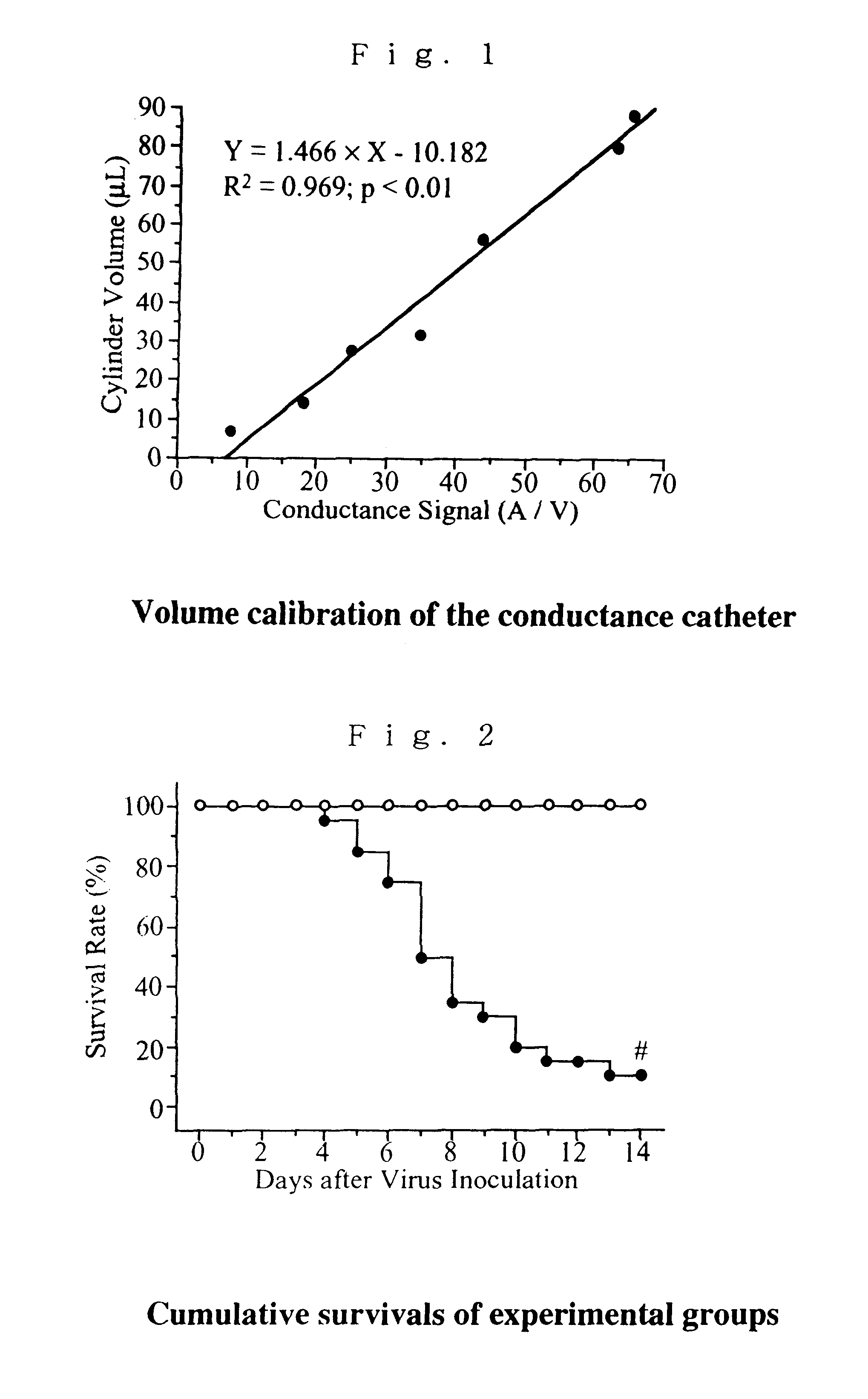
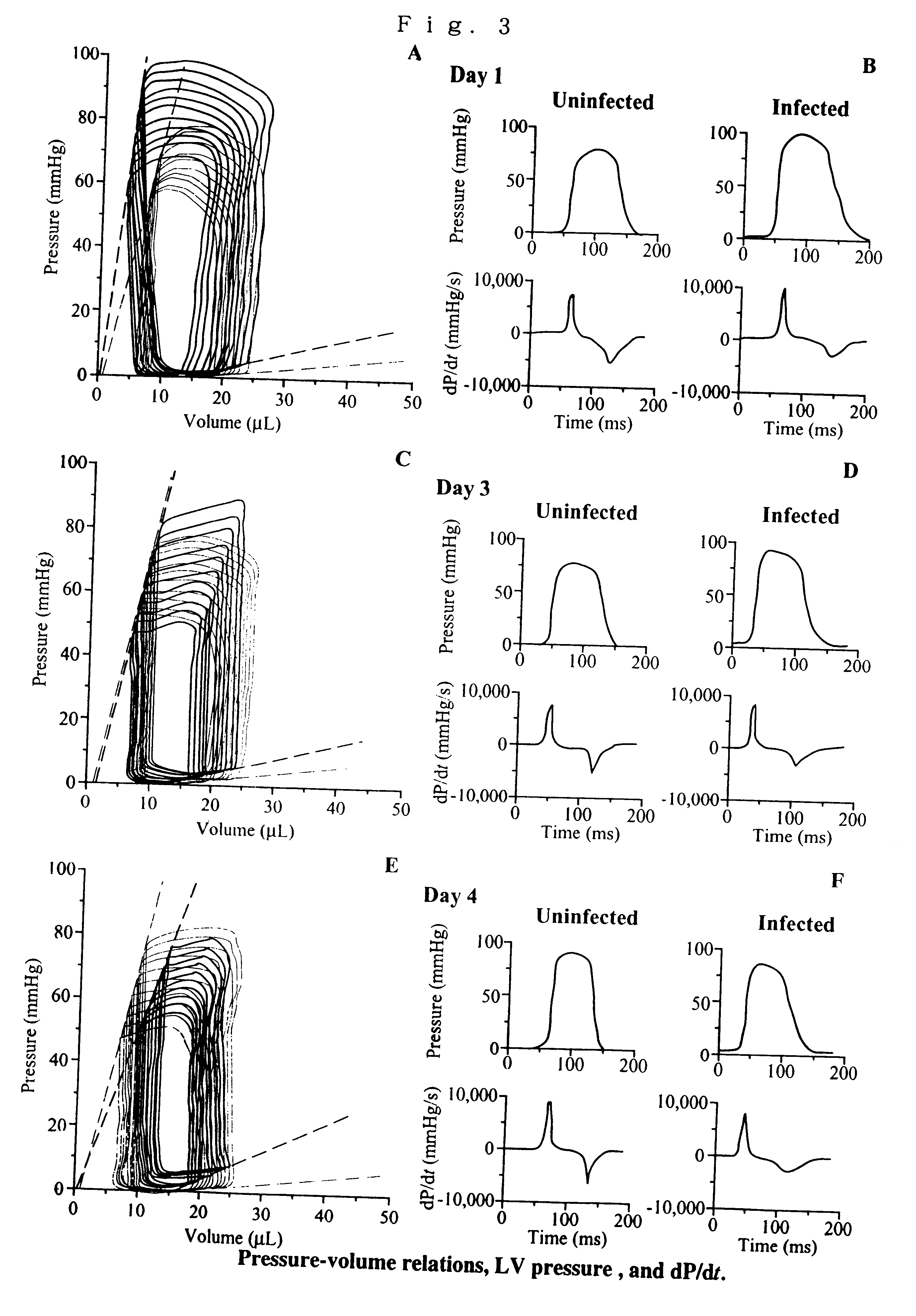
Viral myocarditis is an important cause of congestive heart failure and may lead to dilated cardiomyopathy. However, the hemodynamic changes associated with its acute phase have not been analyzed in detail. This study, performed in a murine model of encephalomyocarditis virus myocarditis, used a new Millar 1.4-Fr conductance-micromanometer system for the in vivo determination of the left ventricular (LV) pressure-volume relationship (PVR).

Methods

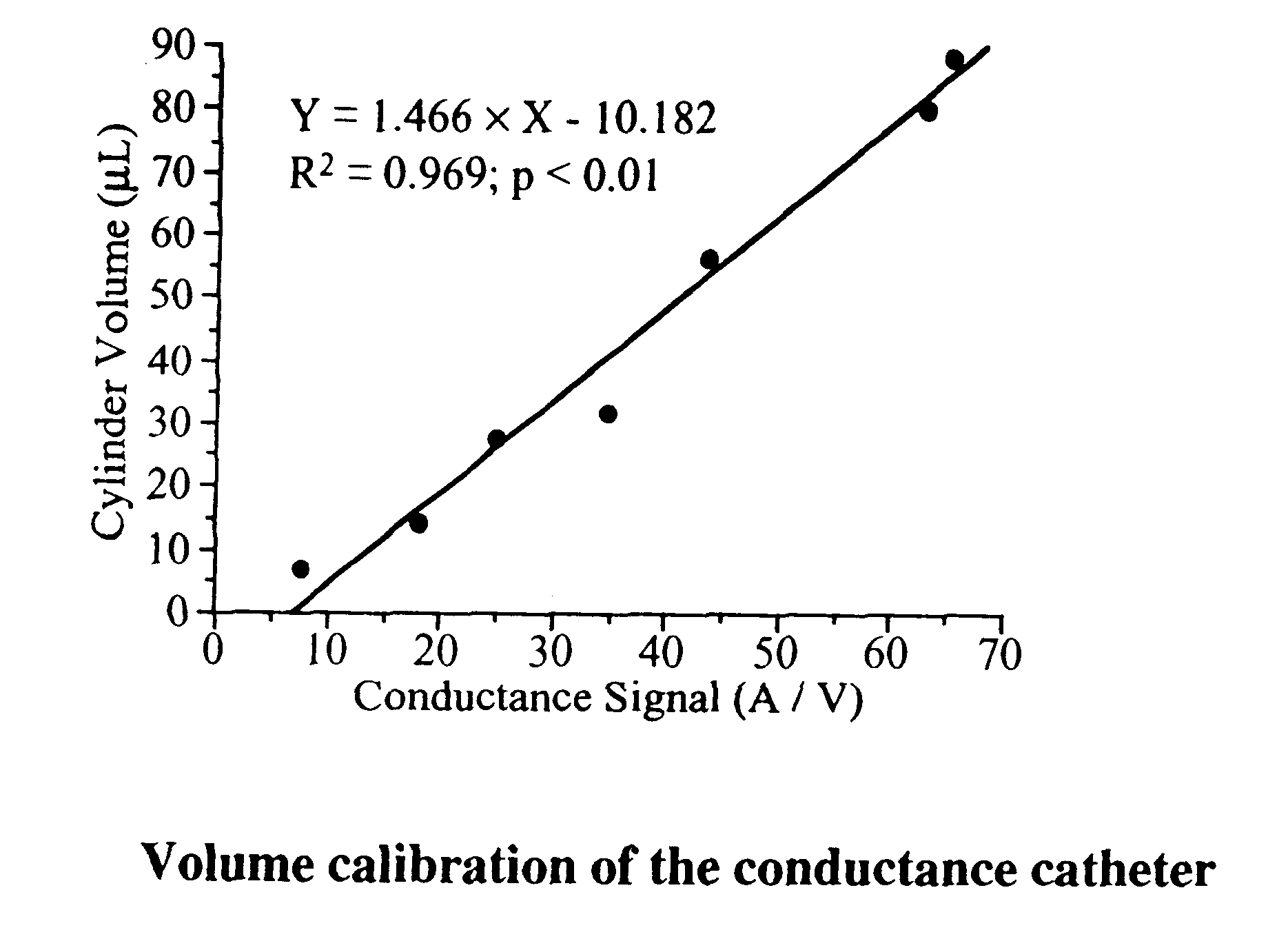
(1) Conductance Catheter System Design

We used the Millar 1.4 Fr catheter (SPR-719, Millar Instruments, Houston, Tex., USA) composed of 4 conductance electrodes and a micromanometer. The distance between the conductance sensor electrodes is 4.5 mm. The conductance system and the pressure transducer controller [Integral 3 (VPR-1002), Unique Medical Co., Tokyo, Japan] were set at a frequency of 20 kHz, the full-scale current selected at 20 .mu.A, and the pressure transducer at 5 .mu.V / V / 100 mmHg. The pressure-volume loops and intracardiac elect...


PUM


Abstract

A method for measuring hemodynamics of an experimental animal enabling repeated measurements, the method comprising the following steps:(i) ligating at least two parts of an artery, between which a part to be incised is located, of the experimental animal, the parts comprising at least one proximal ligated part and at least one distal ligated part, and then incising a part between the ligated parts;(ii) inserting through the incised part a blood pressure-volume simultaneously measuring catheter provided with multiple conductance electrodes and at least one pressure sensor;(iii) loosening the distal ligated part so as to allow an insertion of the catheter and not to cause bleeding in the opened part;(iv) further inserting the catheter so as to introduce the multiple conductance electrodes and the pressure sensor(s) into the heart ventricle; and(v) simultaneously measuring pressure and volume of the heart.

Description

This invention relates to a method for measuring hemodynamics (pressure and volume of the heart) to evaluate pharmacological and therapeutic effects and the like in animal experiments.DESCRIPTION OF RELATED ARTMethods for measuring pressure-volume relationship regarding hemodynamics in animal experiments have been known.In the prior art, especially in a small experimental animal such as a mouse and the like, hemodynamics have been measured by opening the chest of the animal, sticking a measuring instrument such as a needle into the heart of the mouse, and inserting a catheter through the needle. However, since this method is invasive, hemodynamics under physiological conditions cannot be measured.Especially, an experimental animal for genetic manipulation (a mutant) as a model with a serous disease is essentially vulnerable to invasions and tends to easily die. Therefore, there have been disadvantages that for such an experimental animal, it is difficult to used the prior art method...

Claims


Application Information

Patent Timeline
Patent Type & Authority Patents(United States)
IPC IPC(8): A61B5/053A61B5/0215A61B5/0295A61D1/00A61D13/00
CPCA61B5/02028A61B5/0215A61B5/0535A61B5/0538
Inventor NISHIO, RYOSUKEMATSUMORI, AKIRA
Owner NISHIO RYOSUKE
Why Eureka
Try Eureka