Method and a system for determination of somatic cells in milk

a somatic cell and determination system technology, applied in the field of somatic cell determination system, can solve the problems of inability to distinguish between cells and fat particles, high complexity and cost of the instruments used today, and the difficulty of flow cytometry equipmen

Inactive Publication Date: 2004-05-04
CHEMOMETEC AS
View PDF33 Cites 32 Cited by
  • Summary
  • Abstract
  • Description
  • Claims
  • Application Information

AI Technical Summary

Benefits of technology

The physical dimension of an instrument based on the present invention is also such that the instrument will be well suited for transport, thus making it possible for e.g. veterinarians to transport the instrument to or on a location where the analysis is needed. The principle of measurement of the present invention provides a major improvement in the assessment of DNA-containing particles, e.g. somatic cells in a milk or a milk product analyte material, compared to the methods hitherto used for this purpose.
As mentioned above, the sample is preferably at stand still during the exposure. However, in another embodiment, the sample in the domain or sample compartment is moved through the domain or sample compartment during the exposure, and the exposure is performed over a sufficiently short period of time to substantially obtain stand still condition during the exposure. In either case, there is a close control of the volume of the sample from which the exposure is made, which is one very preferred feature of the present invention.

Problems solved by technology

Flow cytometry requires rather elaborate and high cost equipment, partly because of the high accuracy of flow rate necessary to give reliable results, and partly because of the high sensitivity needed to detect the weak signals from the particles in question during the relative short period of time the particle is present in the detector.
Thereby information regarding the particle content is obtained, however distinction between the various particles in the same size distribution, such as distinction between cells and fat particles, is not possible.
Due to the relative high complexity and cost of the instruments used today, most of the assessments of the number of somatic cells in a milk or a milk product analyte are carried out in a laboratory where skilled operators operate the instruments.

Method used


Image

  • Method and a system for determination of somatic cells in milk
  • Method and a system for determination of somatic cells in milk
  • Method and a system for determination of somatic cells in milk

Examples


example 1

Detection of fluorescence signals from Ethidium Bromide (EtBr) bound to DNA in Somatic Cells in Milk at different initial concentration levels of Ethidium Bromide.

The sample material was cow bulk milk. To each of three portions of the same sample material, used for the below Experiments A, B and C, was added a buffer in the ratio of two parts by volume of buffer solution to one part by volume of milk. The buffer solutions were identical, except that they contained different amounts of EtBr. The buffer solutions were prepared according to the guidelines of International IDF standard 148A:1995--"Method C, concerning Flouro-Opto-Electronic Method" (Experiment A, EtBr concentration 33 .mu.g / ml); for Experiment B, the concentration of EtBr was 10% of the prescribed amount, and for Experiment C, it was 1% of the prescribed amount.

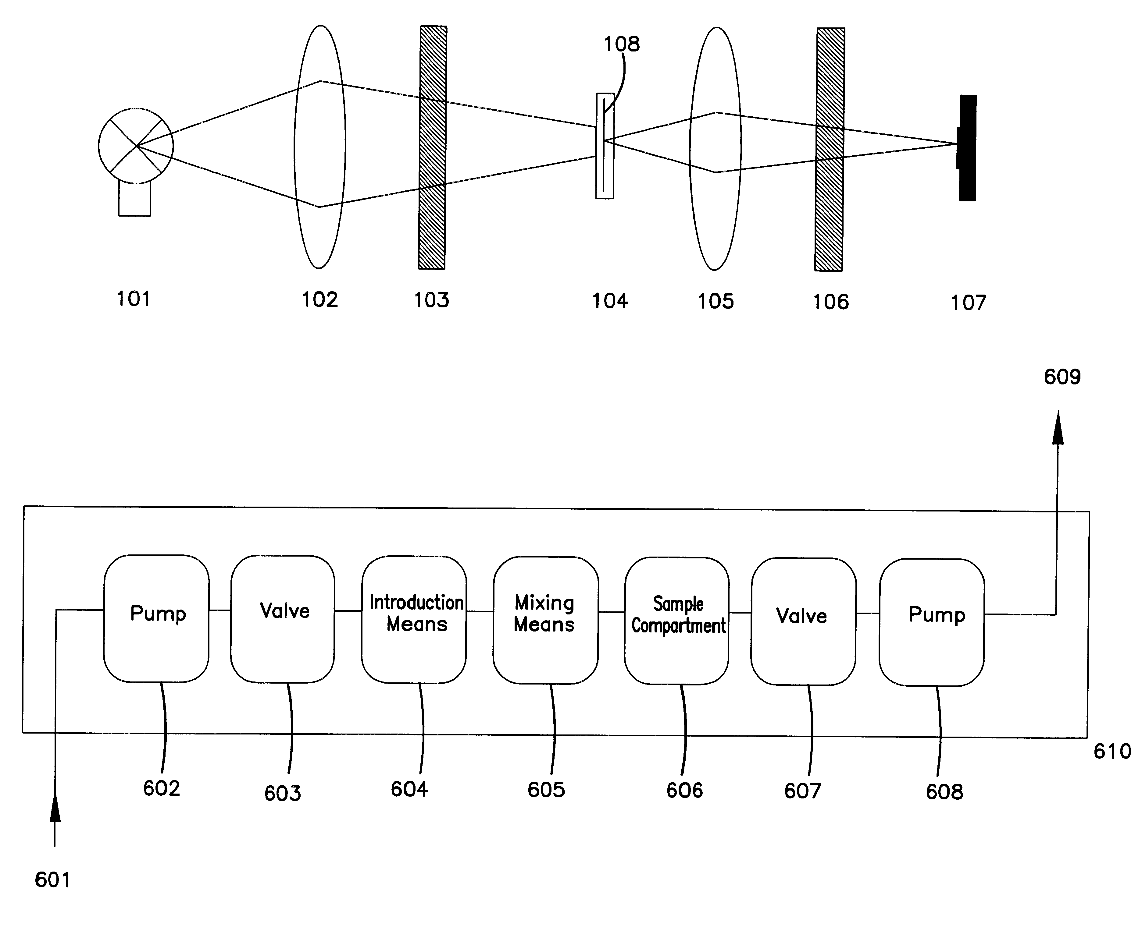
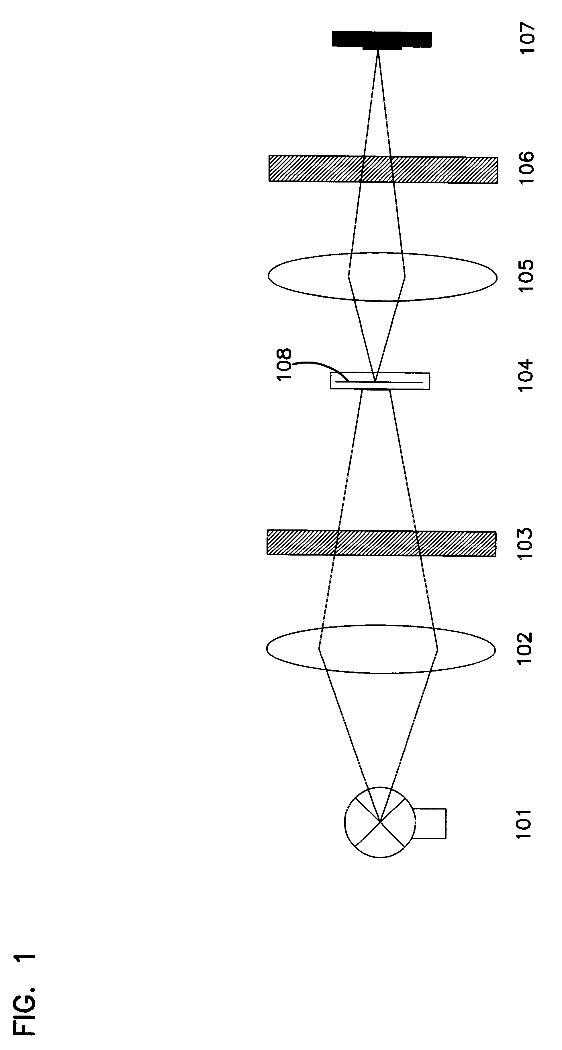
The resulting sample materials were measured in a set-up as follows (cf. FIG. 1): A halogen lamp 101 of type OSRAM (41890 SP 12V,20 W, 10 degree reflector) was u...

example 2

Removal of signal bias by combination of measurements from a linear array of detection elements.

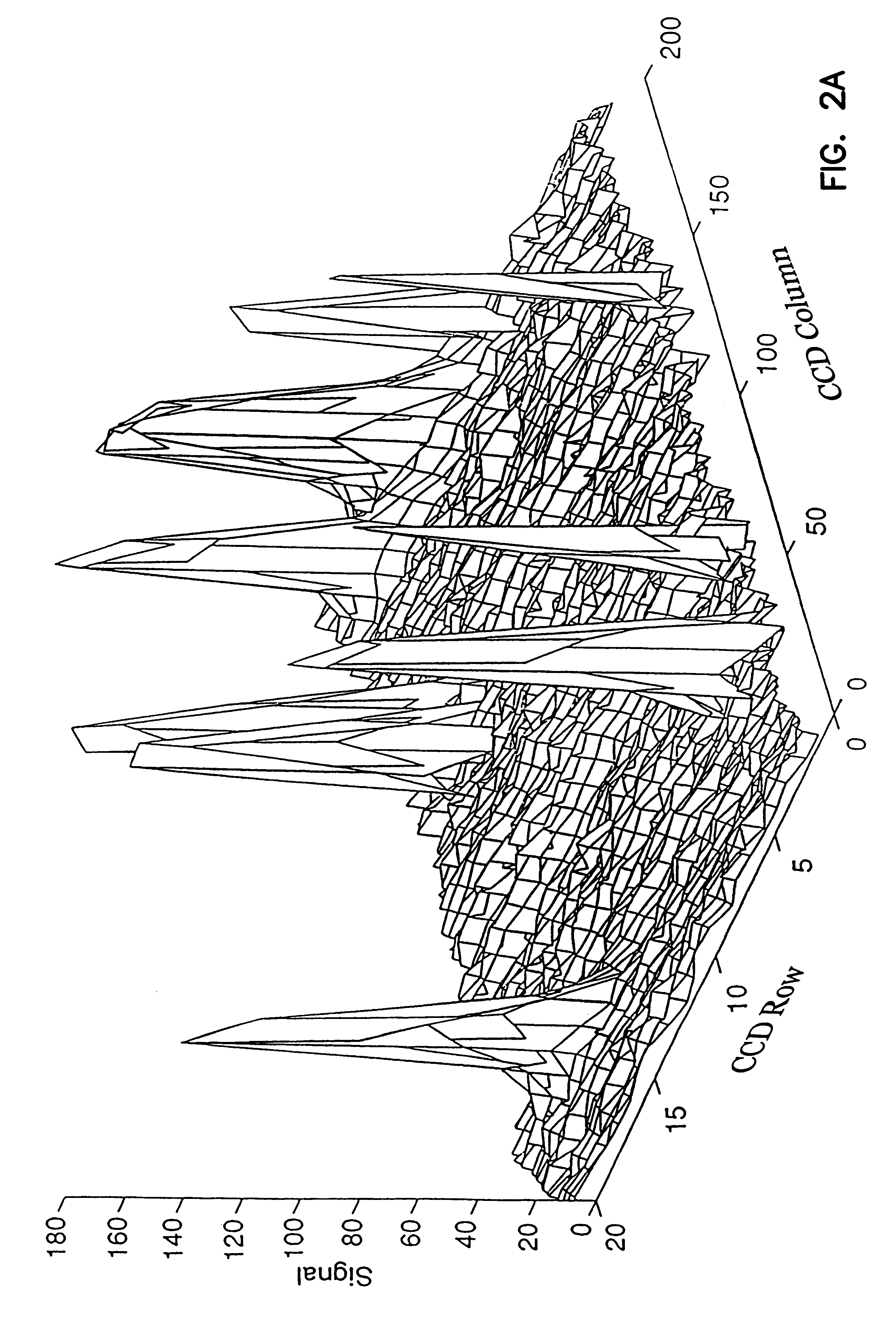
Removal of systematic signal bias can be of interest in the processing of measured signals. In the present example, a linear array of detection elements of the type Hamamatsu (S3902-128Q) was used in an arrangement similar to the one illustrated in FIG. 1. Under the conditions used, the array of detection elements gave a readout which had a systematic bias between detection elements with even index and detection elements with odd index. A series of 2 measurements was carried out using water as sample material.

Results

FIG. 3 shows the results of the measurements of water. FIG. 3A shows the result from the first measurement after the measurement had been adjusted for the mean bias. From FIG. 3A it is apparent that there is a clear difference in the signal intensity of odd and even detection elements, in that elements with an odd index have generally lower signal. FIG. 3B shows the result of ...

example 3

Optical configuration for wide angle collection of signal from a sample

It can be demonstrated that the intensity of any signal collected from a sample is dependent on the square of the collection angle. In conventional automated microscopy, the collection angle is at the most 20 degrees and normally considerably lower, such as 1-5 degrees. Because of the low magnification (or no magnification) which can be used according to the present invention, and the robust processing made possible thereby, a much larger collecting angle can be used. In the present example, two different optical arrangements are used to obtain a collection angle of approximately 40 and approximately 70 degrees, respectively.

FIG. 4 illustrates an optical arrangement which produces a collection angle of approximately 40 degrees when collecting a signal from a sample compartment 401 and projecting it onto detection elements 404, by using two achromatic lenses, one 402 of the type Melles Griot 01 (LAO 014: F=21 mm, ...


PUM

PropertyMeasurementUnit
volumeaaaaaaaaaa
thicknessaaaaaaaaaa
thicknessaaaaaaaaaa

Abstract

Assessment of the number of somatic cells in a liquid milk or milk product analyte material comprises arranging a volume of a liquid sample representing the analyte material in a sample compartment and allowing electromagnetic signals from the sample in the compartment to pass through and be exposed to the exterior. There is exposed onto an array of active detection elements, an at least one-dimensional spatial representation of electromagnetic signals from the sample. The representation is detectable as an intensity by individual active detection elements. Representations of electromagnetic signs from the somatic cells are identified. The size of the volume of the liquid sample is sufficiently large to permit the assessment of the number of somatic cells to fulfill a predetermined requirement to the statistically quality of the assessment based on substantially one exposure.

Description

This invention relates to a method and a system for the determination or assessment of the number of somatic cells (or fragments thereof, the fragments to be understood to be included whenever somatic cells are mentioned in the following) in a milk or a milk product analyte material. The present invention relates to the assessment of somatic cells in milk product analyte such as, raw milk collected at cow side, raw milk collected during milking, bulk milk delivered by the dairy farmer, milk and milk products produced by dairies and milk samples being measured in central laboratories.DESCRIPTION OF THE RELATED ARTDeterminations or assessments of the number of somatic cells in a milk or a milk product analyte have been performed by various methods. One of these methods is flow cytometry; instrument for performing flow cytometry is available, e.g., from Becton, Dickinson and Company, Franklin Lakes, USA.For example, EP 0 556 971 describes a flow cytometric method for assessing the numb...

Claims


Application Information

Patent Timeline
Patent Type & Authority Patents(United States)
IPC IPC(8): G01N27/00G01R27/00G01R27/32G01R27/04
CPCG01N15/1463G01N21/51G01N21/6486G01N33/04G01N21/59G01N2015/1486
Inventor HANSEN, FRANS EJNER RVANGLENSBJERG, MARTINARNVIDARSON, BORKURJEPPESEN, JESPER MYRON
Owner CHEMOMETEC AS
Why Eureka
Try Eureka