Method of fast and reliable tissue differentiation using diffusion-weighted magnetic resonance imaging

a tissue differentiation and diffusion-weighted technology, applied in the field of tissue differentiation fast and reliable using diffusion-weighted magnetic resonance imaging, can solve the problems of not being completely successful in determining tumor margins using this "conventional" methodology, the mono-exponential signal decay versus b-factor relationship just mentioned may not be necessarily accurate, and the method, even when used in conjunction with contrast enhanced t1 and t2 relaxation-weighted imaging, is not totally successful

Inactive Publication Date: 2004-06-15
THE BRIGHAM & WOMEN S HOSPITAL INC
View PDF7 Cites 32 Cited by
  • Summary
  • Abstract
  • Description
  • Claims
  • Application Information

AI Technical Summary

Problems solved by technology

Nevertheless, the determination of tumor margins using this "conventional" methodology still is not entirely successful.
Again, however, these methods, even when used in conjunction with contrast enhanced T1 and T2 relaxation-weighted imaging, are not totally successful.
In practice in clinical magnetic resonance systems, however, the gradients typically are applied for a moderate duration of several tens of milliseconds, and the leading edges of the respective bursts are separated by delays of a similar length of time.
Still other experiments, however, have suggested that the mono-exponential signal decay versus b-factor relationship just mentioned may not be necessarily accurate.
This model, however, has been criticized.
This, however, was known to be technically incorrect for the reasons discussed above.
However, it also has been recognized that this is not believed to be a totally technically correct interpretation of the various diffusion phenomena under analysis.
It has been found that these contrast agents do not enhance the magnetic resonance signal strength for all tumor types.
However, these contrast agents also can add complexity to, or even ruin, the image analyses they are intended to enhance.
Nevertheless, EPI has been shown to be susceptible to ghosting artifacts and image distortion, among other problems.

Method used


Image

  • Method of fast and reliable tissue differentiation using diffusion-weighted magnetic resonance imaging
  • Method of fast and reliable tissue differentiation using diffusion-weighted magnetic resonance imaging
  • Method of fast and reliable tissue differentiation using diffusion-weighted magnetic resonance imaging

Examples


Embodiment Construction

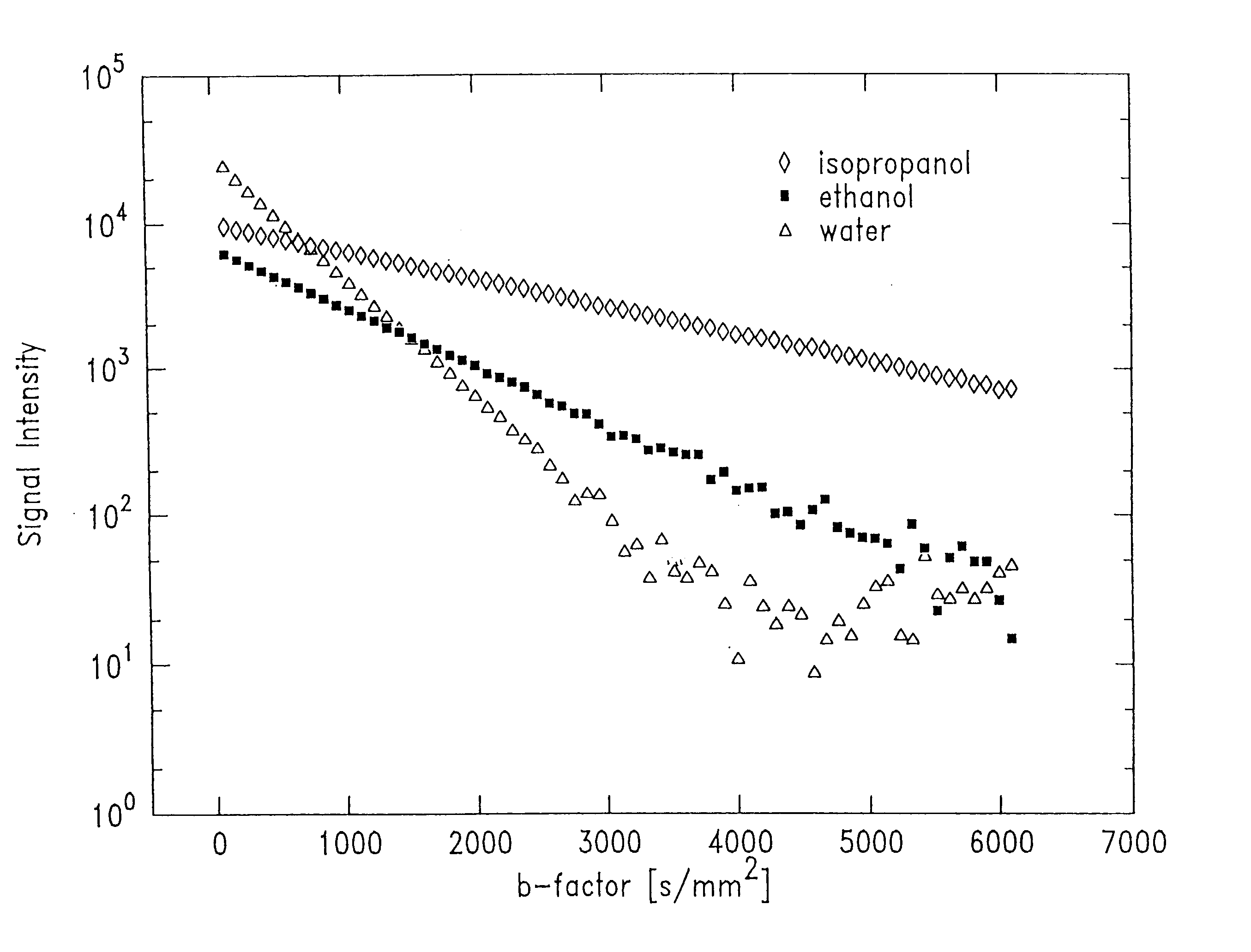
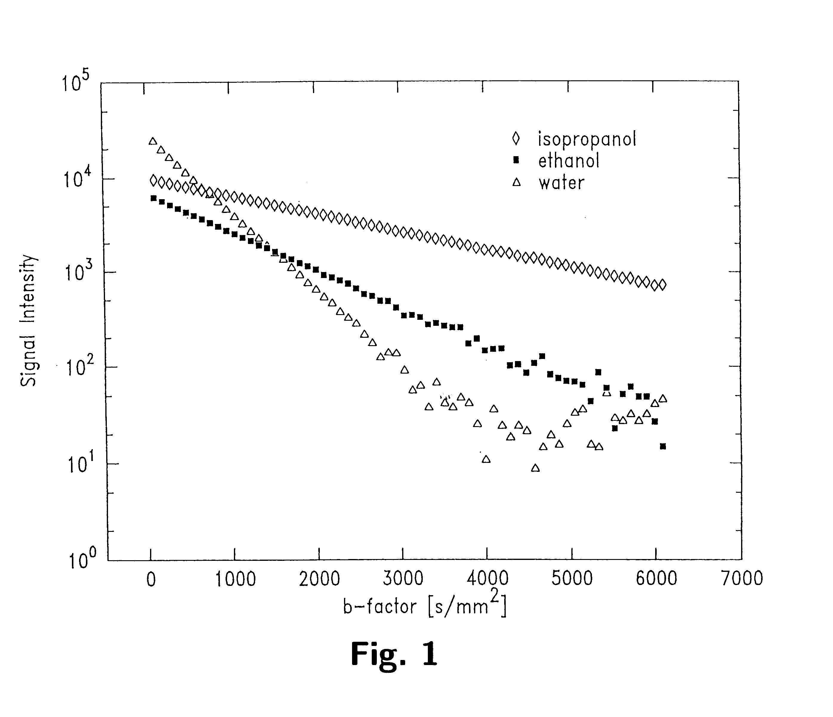
With wide b-factor range diffusion scans along a single column it has been demonstrated that human brain water attenuation is better described with a bi-exponential model than a mono-exponential model as previously believed. Thus, data in image formats was obtained with line scan diffusion imaging (LSDI). Specifically, 15 brain tumor scans were performed on patients. One patient was studied both before and after contrast agent administration. Altogether, four examinations were carried out before contrast administration. The data relating to one patient had to be discarded because of motion artifacts. The pathologies of the remaining 13 patients included 8 glioblastoma multiforme, 2 astrocytomas and 3 metastases. In addition, one normal patient and one stoke patient two days after the onset of symptoms also were scanned.

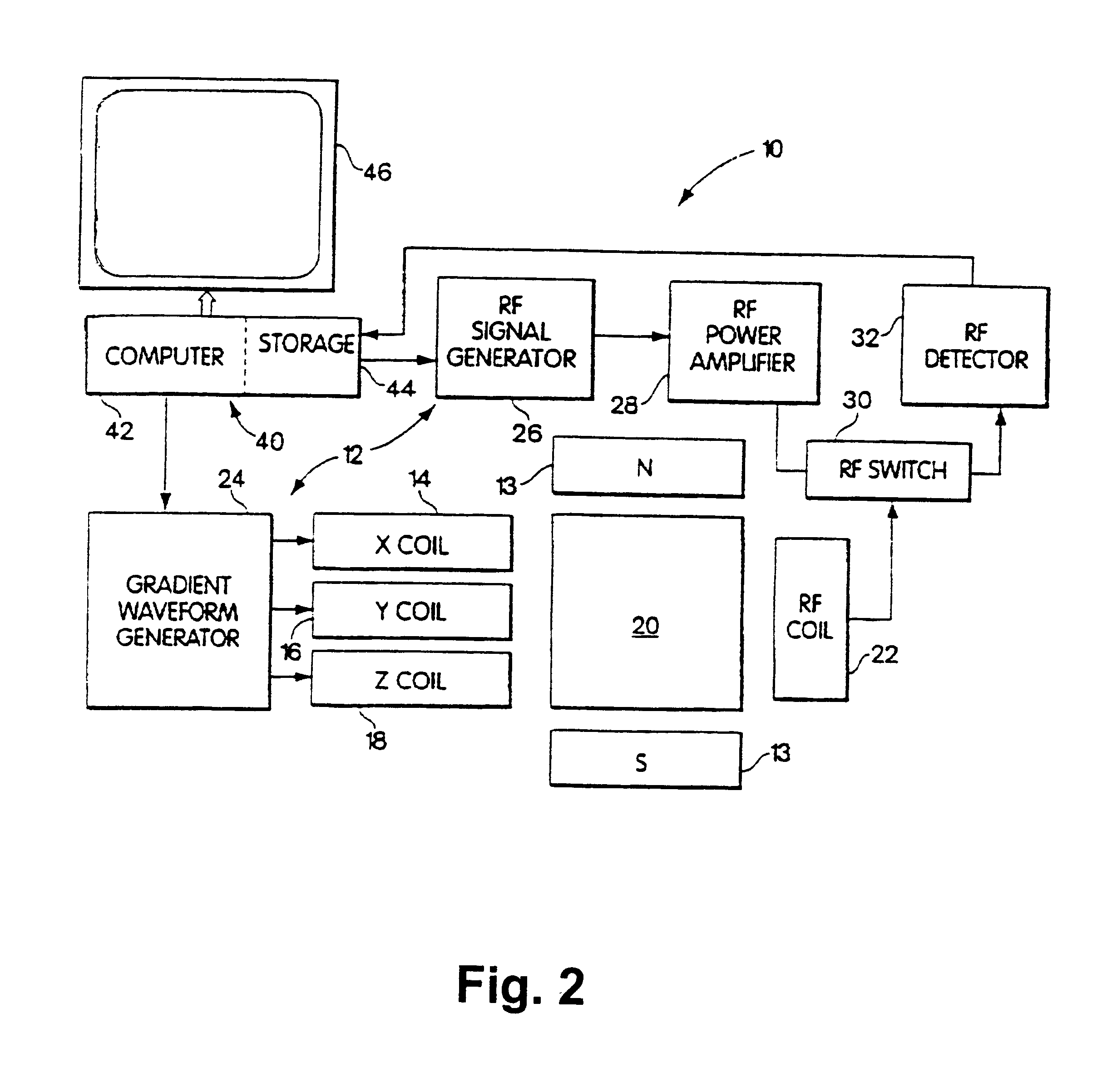
A wide range b-factor LSDI sequence and protocol was implemented on a 1.5 Tesla Horizon Echospeed (GE Medical Systems, Milwaukee, Wis.) system with software release 5...


PUM


Abstract

Quantified differences, such as chi<2 >error parameters, between a mono-exponential, logarithmic best fit of a series of line scan diffusion-weighted magnetic resonance signals taken over a range of b-factors between about 100 and about 5000 sec / mm<2 >are obtained. The quantified differences so generated are displayed as an image wherein the brightness of each pixel depends upon the size of its associated quantified difference. The resulting image is characterized by high signal to noise ratio and distinctness between varying tissue types.

Description

1. Field of InventionThe invention generally relates to methods for obtaining, processing and displaying parameters associated with in vivo tissue water diffusion as pathologically significant images using magnetic resonance imaging. More particularly, the invention relates to methods for obtaining and processing diffusion weighted output signals from a magnetic resonance imaging apparatus, and to the fast creation of high definition images of internal bodily tissue(s) utilizing the so processed magnetic resonance output signals.2. Summary of the Prior ArtTissue differentiation and localization always have been basic goals of magnetic resonance imaging. Indeed, the desire to distinguish between normal tissue and tumor tissue using magnetic resonance imaging techniques was recognized at least thirty years ago. At that time, it was realized that the spin-lattice, so-called "T1", as well as the spin-spin, so-called "T2", relaxation parameters are different between normal and cancerous ...

Claims


Application Information

Patent Timeline
Patent Type & Authority Patents(United States)
IPC IPC(8): G01R33/54G01R33/56A61B5/055G01R33/563
CPCG01R33/56A61B5/055G01R33/5608G01R33/56341
Inventor MAIER, STEPHAN E.MULKERN, JR., ROBERT V.
Owner THE BRIGHAM & WOMEN S HOSPITAL INC
Why Eureka
Try Eureka