
Method for chemiluminescent detection

a chemiluminescent and detection method technology, applied in biochemistry apparatus, biochemistry apparatus and processes, photographic processes, etc., can solve the problems of increasing the number of reagents, requiring more time to complete the assay, and many steps

Inactive Publication Date: 2004-07-20
EMP BIOTECH
View PDF6 Cites 5 Cited by
  • Summary
  • Abstract
  • Description
  • Claims
  • Application Information

AI Technical Summary

Benefits of technology

This method enhances signal amplification, reduces assay time and costs, and allows for the reuse of analytes, improving the sensitivity and efficiency of nucleic acid and immunoassays by using a non-enzymatic, triggerable chemiluminescent compound formation.

Problems solved by technology

However, a disadvantage of such methods as they are currently practiced in the field is that many steps are required in the assay protocol, requiring more time to complete the assay.
Moreover, a greater number of reagents are required which means greater cost.
Most commercially available chemiluminescent detection systems employ enzyme conjugates to increase detection sensitivity through amplification of the signal and, therefore, suffer from the same disadvantages described above.
The disadvantage of this format is that, because the analyte must be subjected to heat and / or base, it is not further utilized for additional testing and analysis.
One disadvantage of this assay is that, because it is enzyme-based, the activity of the enzyme, its stability and its shelf-life need to be monitored as discussed above.
Because heating a caustic solution of chemical base to at or near boiling temperatures would be both dangerous and impractical, this prior application teaches the use of an activating film that may contain a solid chemical component immobilized on or impregnated therewith, which when acted upon by an energy source, such as heat, releases an activating substance capable of decomposing a triggerable chemiluminescent compound to produce a chemiluminescent signal for the detection of a target molecule.

Method used


Image

  • Method for chemiluminescent detection
  • Method for chemiluminescent detection
  • Method for chemiluminescent detection

Examples


example 2

Dot Blot Hybridization of Methylene Blue-Labeled Oligonucleotide to Target DNA

Following PCR amplification of the target DNA, said DNA was spotted on a Hybond+nylon membrane (Amersham Biosciences Corporation), along with negative controls of linearized pUC19 DNA at various concentrations ranging from 25 to 500fmoles in a total volume of 1 microliter. Spots were allowed to dry. The DNA was subsequently denatured and fixed as follows: 1 minute soak in 1.5 M NaCl; 0.5 M NaOH, followed by fixation by baking at 120.degree. C. for 40 minutes, followed by 5 minute soak in 1.5 M NaCl; 0.5 M Tris-Cl pH 7.5. Hybridization was as follows: The filter membrane was soaked in prehybridization buffer (0.25 M Na--PO.sub.4 ; pH 7.2; 7% (w / v) SDS for 45 minutes at 40.degree. C. in a total volume of 0.5 ml per cm.sup.2 / membrane. The labeled oligonucleotide probe was added directly to the prehybridization buffer at a final concentration of about 2 .mu.moles / ml and incubated for 16 h at 40.degree. C. The...

example 3

Preparation of the Inventive Film

Ten mg of an appropriate chemiluminescent olefin are dissolved in 100 ml (0.37 mM) of n-hexane or methanol. A Hybond-N neutral nylon membrane (Amersham Biosciences Corporation) was dipped in the olefin solution and allowed to air dry.

example 4

Formation of a Triggerable Chemiluminescent Compound on the Inventive Film

Hybridization was detected by first assembling the sandwich formation shown in FIG. 6A. In order to detect a signal, a membrane containing hybridized target DNA from Example 2 is placed (DNA side up) on a glass plate. The inventive membrane with chemiluminescent precursor immobilized therewith was subsequently placed on top of the membrane with the target molecule. A sheet of non-transparent, black paper is placed over the inventive film and another glass plate was placed on top of the whole sandwich formation. The sandwich formation was then exposed to red light for 15 minutes by using an appropriate cut-off filter and irradiating through the glass plate containing the hybridized target DNA. This allows for formation of a triggerable chemiluminescent precursor compound on the inventive film, which may be subsequently triggered as described in Example 5 below.


PUM

PropertyMeasurementUnit
wavelengthaaaaaaaaaa
wavelength rangeaaaaaaaaaa
wavelengthaaaaaaaaaa

Abstract

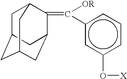
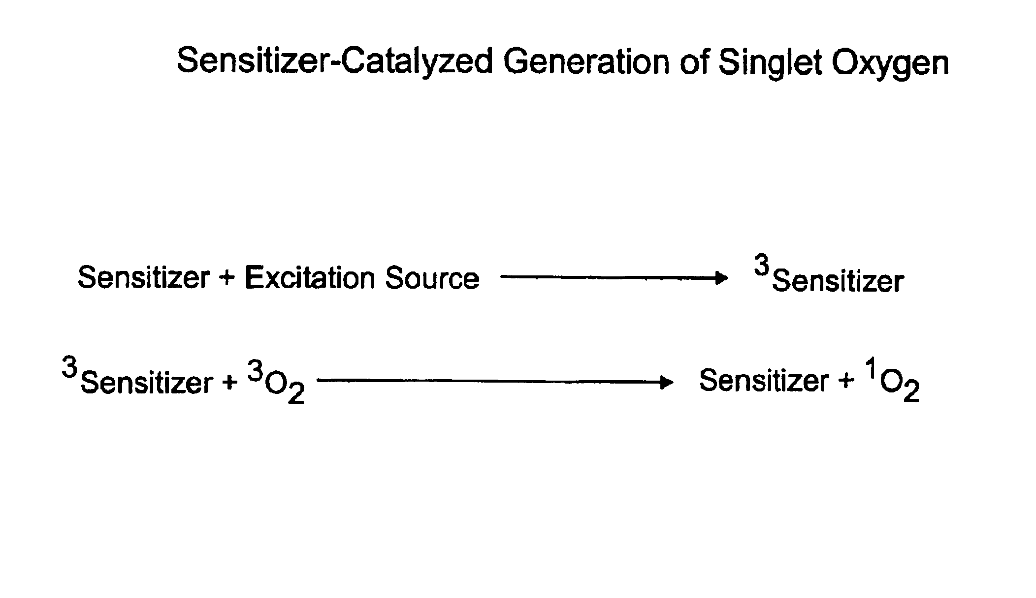
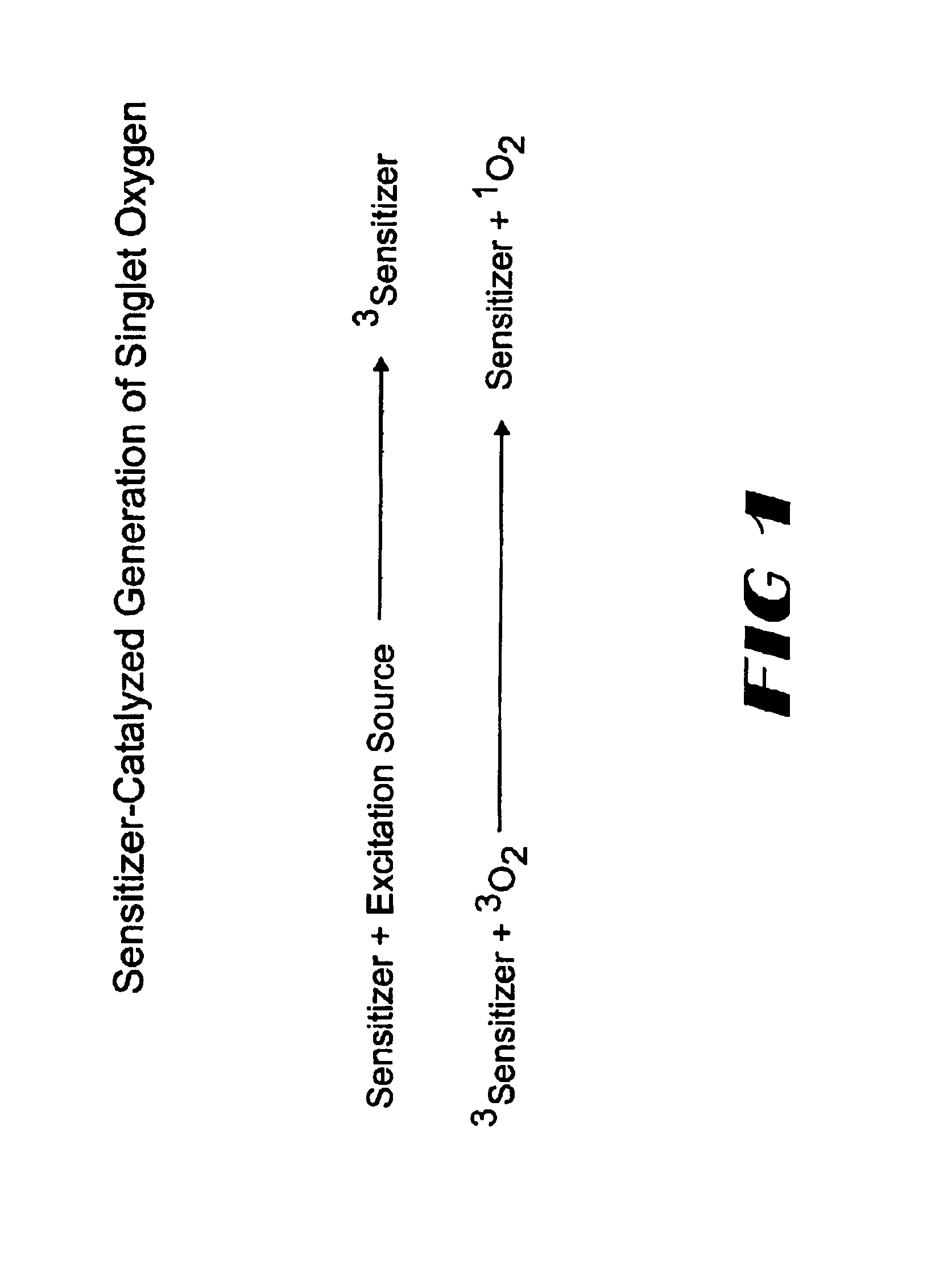
The invention provides chemiluminescent assays that incorporate a film including at least one chemiluminescent precursor immobilized therewith which produces a triggerable chemiluminescent compound, the film being free of compounds which generate singlet oxygen and being adapted for use with a sensitizer-labeled agent or agent probative of the analyte.

Description

The present invention relates to chemiluminescent assays that incorporate a film which includes a chemiluminescent precursor component immobilized therewith which produces a triggerable chemiluminescent compound for detection of a target molecule.BACKGROUND OF RELATED TECHNOLOGYRecently a variety of non-isotopic labeling methods have been developed to replace radioactive labels in DNA probe-based assays. It is most common in such methods to use marker enzymes to detect nucleic acid probes using either colormetric, chemiluminescent, bioluminescent or fluorescent methods. Each of these methods have been used reliably for both hybridization of DNA probe-based assays for nucleic acid detection as well as in solid-phase immunochemical assays wherein the target molecule is typically an antigen of interest.Regardless of the type of non-isotopic detection method used, the labels are typically measured directly with fluorophores (without use of enzymes) or indirectly using enzyme amplificati...

Claims


Application Information

Patent Timeline
Patent Type & Authority Patents(United States)
IPC IPC(8): C12Q1/00C12M3/00C12Q1/28C12Q1/06C12Q1/08C12Q1/42C12M1/34G01N33/53G03C5/04C09K11/07C12QC12Q1/04C12Q1/68G01N33/543G01N33/558G01N33/569
CPCC09K11/07C12Q1/04C12Q1/6816G01N33/543G01N33/558Y10S435/975Y10S435/968
Inventor LEVISON, DEREK W. K.MOLLER, UWELEVISON, STUART
Owner EMP BIOTECH
Learn More