Methods and devices for blood vessel harvesting

a technology for harvesting and veins, applied in the field of endoscopic surgery, can solve the problems of long incisions created in the leg, slow healing of long incisions, and inability to remove veins, and achieve the effects of reducing complications, facilitating vein pulling, and reducing incisions

Inactive Publication Date: 2005-01-11
GEN SURGICAL INNOVATIONS
View PDF33 Cites 146 Cited by
  • Summary
  • Abstract
  • Description
  • Claims
  • Application Information

AI Technical Summary

Benefits of technology

The methods and devices presented herein allow surgeons to harvest veins without making the long incisions previously required. The procedure is a minimally invasive procedure that requires just a few small incisions, one at either end of the saphenous vein. The procedure is accomplished with endoscopic or laparoscopic instruments under the guidance of an endoscope. The surgeon makes one small incision at each end of the saphenous vein. After making the incisions, the surgeon inserts a tunneling instrument or blunt dissector into one incision and advances or pushes along the saphenous vein to make a small tunnel along the saphenous vein. The surgeon then inserts the long balloon described below in the tunnel and inflates the balloon to enlarge and further propagate the tunnel. The surgeon may use the balloon to dissect the fat and skin overlying the saphenous vein away from the vein, and also to enlarge the tunnel to an appropriate size. When the tunneling is completed, the surgeon removes the balloon and seals the tunnel at both ends. The surgeon then injects carbon dioxide, nitrous oxide, or other suitable gas into the tunnel at sufficient pressure (typically 5-15 mmHg) to inflate the tunnel and create room for laparoscopic instruments. The surgeon then inserts an endoscope or laparoscope through the seal to provide a view of the procedure, and inserts an endoscopic vein harvesting device described in detail below. With the vein harvester in place, the surgeon manipulates the vein harvester to strip the connective tissue from the vein, identify and cut side branches, and remove the vein from its channel in the leg. After the vein is loosened or dissected free from its channel in the leg, the surgeon can cut the proximal and distal ends and easily pull the vein from the leg. The small skin incisions are stitched so they may heal. The small incisions heal much more readily, with fewer complications and far less pain, than the operation now in use.
The surgeon operating with the vein harvesting device has a choice of tunneling instruments. A typical blunt dissection is performed with a long stiff rod with a round tip, and the tip may be enlarged. The surgeon may also use a device referred to as a peanut, which is long rod with a soft fabric or foam tip. These devices are used to create a small tunnel, and then the balloon dissector may be inserted to enlarge the tunnel. The balloon dissector described below allows the tunneling operation to be accomplished by the balloon, thus allowing easier and less traumatic creation of the initial tunnel, and also permitting the surgeon to create the enlarged tunnel with this single device. The device may be used as an alternative to the other methods of enlarging the tunnel.

Problems solved by technology

Coronary bypass surgery is required when these other methods of treatment cannot be used or have failed to clear the blocked artery.
As can be seen from the description of the harvesting operation, the harvesting operation is very traumatic in its own right.
The vein harvesting operation is often the most troublesome part of the operation.
The long incisions created in the leg can be slow to heal and very painful.
Complications resulting from the vein harvesting operation can also hinder the patient's recovery from the entire operation.

Method used


Image

  • Methods and devices for blood vessel harvesting
  • Methods and devices for blood vessel harvesting
  • Methods and devices for blood vessel harvesting

Examples


Embodiment Construction

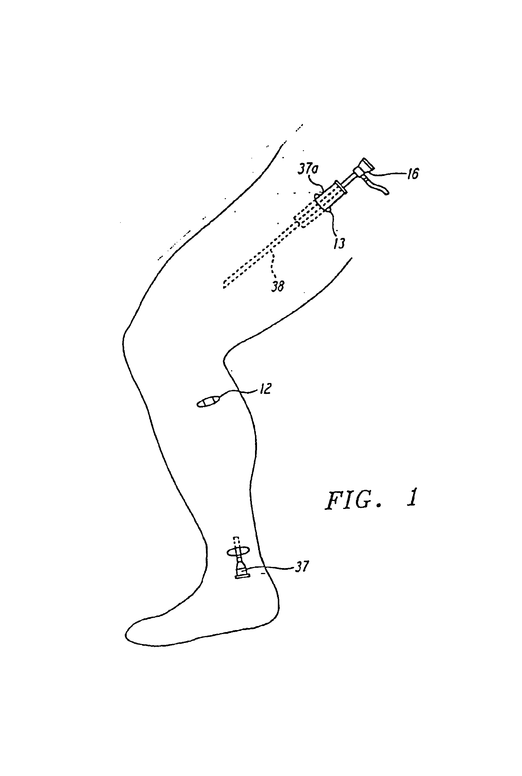
The methods and devices presented herein take advantage of laparoscopic procedures to lessen the trauma of vein harvesting operations. Instead of making an incision along or over the entire length, or essentially the entire length of the vein to be harvested, the procedure may be conducted with only a few small incisions. All that is needed is a working space large enough to allow the surgeon to use the tool and view the operation through a laparoscope. In the preferred embodiment of the method, the surgeon creates a working space under the skin and over the saphenous vein using laparoscopic techniques. The surgeon makes several small incisions to expose the saphenous vein. These incisions are referred to as cut-downs. A distal incision near the knee and a proximal incision at the groin are preferred. If the entire length of the saphenous vein is to be harvested, an additional incision can be made close to the ankle. The saphenous vein can be seen through the cut-downs. It will be a...


PUM


Abstract

Methods and devices for harvesting veins from the body. The methods and devices allow vein harvesting using laparoscopic procedures. A working space is created over the vein using standard laparoscopic procedures and a side-hooked wire is inserted into the working space and twisted to insert the side-hook under the vein. The wire is then pulled or drawn along the vein to separate the vein from the surrounding tissue. In an alternative embodiment, vein separation is accomplished by threading a soft rubber tube under the vein, grasping the ends of the tube so as to surround the vein, and then pulling the tube along the vein. In other embodiments, the working space is created with everting balloons.

Description

FIELD OF THE INVENTIONThis invention relates to methods and devices for endoscopic surgery, in particular to methods and devices for dissecting tissue to create a working space for endoscopic instruments.BACKGROUND OF THE INVENTIONNumerous surgical procedures have been developed to replace arteries that have become blocked by disease. The aortocoronary bypass surgery is perhaps the most important of these bypass operations. The coronary arteries supply blood to the heart. As a result of aging and disease, coronary arteries may become blocked by plaque deposits, stenosis, or cholesterol. In some instances, these blockages can be treated with artherectomy, angioplasty or stent placement, and coronary bypass surgery is not required. Coronary bypass surgery is required when these other methods of treatment cannot be used or have failed to clear the blocked artery. In the coronary bypass surgery, a vein is harvested from elsewhere in the body and grafted into place between the aorta and ...

Claims


Application Information

Patent Timeline
Patent Type & Authority Patents(United States)
IPC IPC(8): A61B17/00A61B17/32A61B17/34A61B17/11
CPCA61B17/00008A61B2017/00557A61B2017/3419A61B2017/320044A61B2017/00969
Inventor FOGARTY, THOMAS J.MOLLENAUER, KENNETH H.MONFORT, MICHELLE Y.HERMANN, GEORGE D.WILL, ALLAN R.
Owner GEN SURGICAL INNOVATIONS
Why Eureka
Try Eureka