
Method, device and computer program for determining the blood flow in a tissue or organ region

a tissue or organ region and blood flow technology, applied in the field of methods, devices and computer programs for determining the blood flow in tissue or organ regions, can solve the problems of not being able to confin the tissue or organ region to a specific region, and the known methods are not suitable for determining the regional blood flow, so as to achieve the effect of determining with eas

Inactive Publication Date: 2005-02-08
RENEW GRP +1
View PDF12 Cites 44 Cited by
  • Summary
  • Abstract
  • Description
  • Claims
  • Application Information

AI Technical Summary

Benefits of technology

The object forming the basis of the invention is to provide a method, a device and a computer program, with the aid of which the blood flow in a precisely defined, possibly also extended, tissue or organ region can be determined with ease during routine application in the operating theatre.
The efficiency of modern computers makes it possible to determine the appropriate transport function g(t), which makes the said term a minimum, within a very short time.
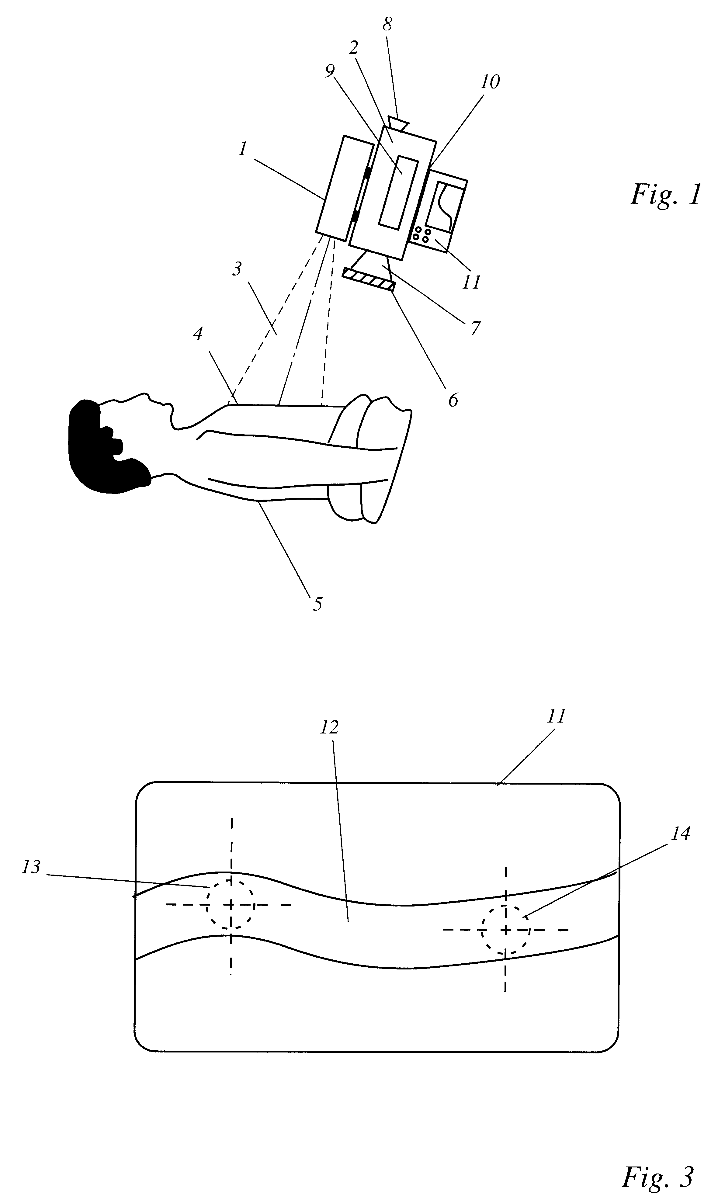
According to an advantageous embodiment, the detector unit is an imaging unit, which enables at least one image area to be defined as a measurement region and at least one image area to be defined as a reference region. To determine the fluorescence curve, more precisely the fluorescence curves, only the defined areas are used. The fluorescence curve of the measurement region is correlated with the fluorescence curve of the reference region. Random disturbances such as fluctuations in chromophore concentration, for example, can be eliminated during calculation of the blood flow by forming the difference or the relation of the two curves.
According to an advantageous further development of the computer program, this has a program module which enables the selection of at least one measurement region and at least one reference region within the image recorded by the video camera. For evaluation, the fluorescence curve of the measurement region is correlated with the fluorescence curve of the reference region. Random disturbances such as fluctuations in chromophore concentration, for example, can be eliminated during calculation of the blood flow by forming the difference or the relation of the two curves.

Problems solved by technology

The known methods are not suitable for determining the regional blood flow quantitatively on the basis of the fluorescence signal.
Moreover, these methods are not suitable for confining the tissue or organ region to be examined to a precisely defined area.

Method used


Image

  • Method, device and computer program for determining the blood flow in a tissue or organ region
  • Method, device and computer program for determining the blood flow in a tissue or organ region
  • Method, device and computer program for determining the blood flow in a tissue or organ region

Examples


Embodiment Construction

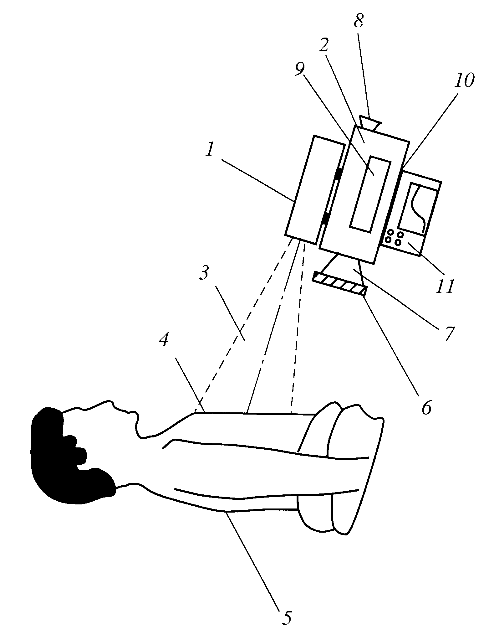
Referring to the drawings, FIG. 1 schematically shows a device according to the invention in use during an operation in a view not to scale. A safety case 1, into which an infrared laser light source with a peak emission of 780 nm is integrated, together with a CCD camera 2 and an arithmetic or evaluator unit 11, forms a compact unit, which can be carried and operated with one hand, has an accumulator and is therefore usable independently of mains power.

The expanded laser light 3 discharging from the safety case 1 has a surface intensity of less than 1 mW / cm2 and lies below the limit value of the maximum permitted irradiation of the cornea of the eye, and as a result no safety glasses need to be worn in the vicinity of the device.

The expanded beam bundle 3 of the infrared laser light source irradiates the approximately 30 cm wide field of operation 4, which is located approximately at a distance of 70 cm from the safety case 1. Indocyanine green previously administered to the patien...


PUM


Abstract

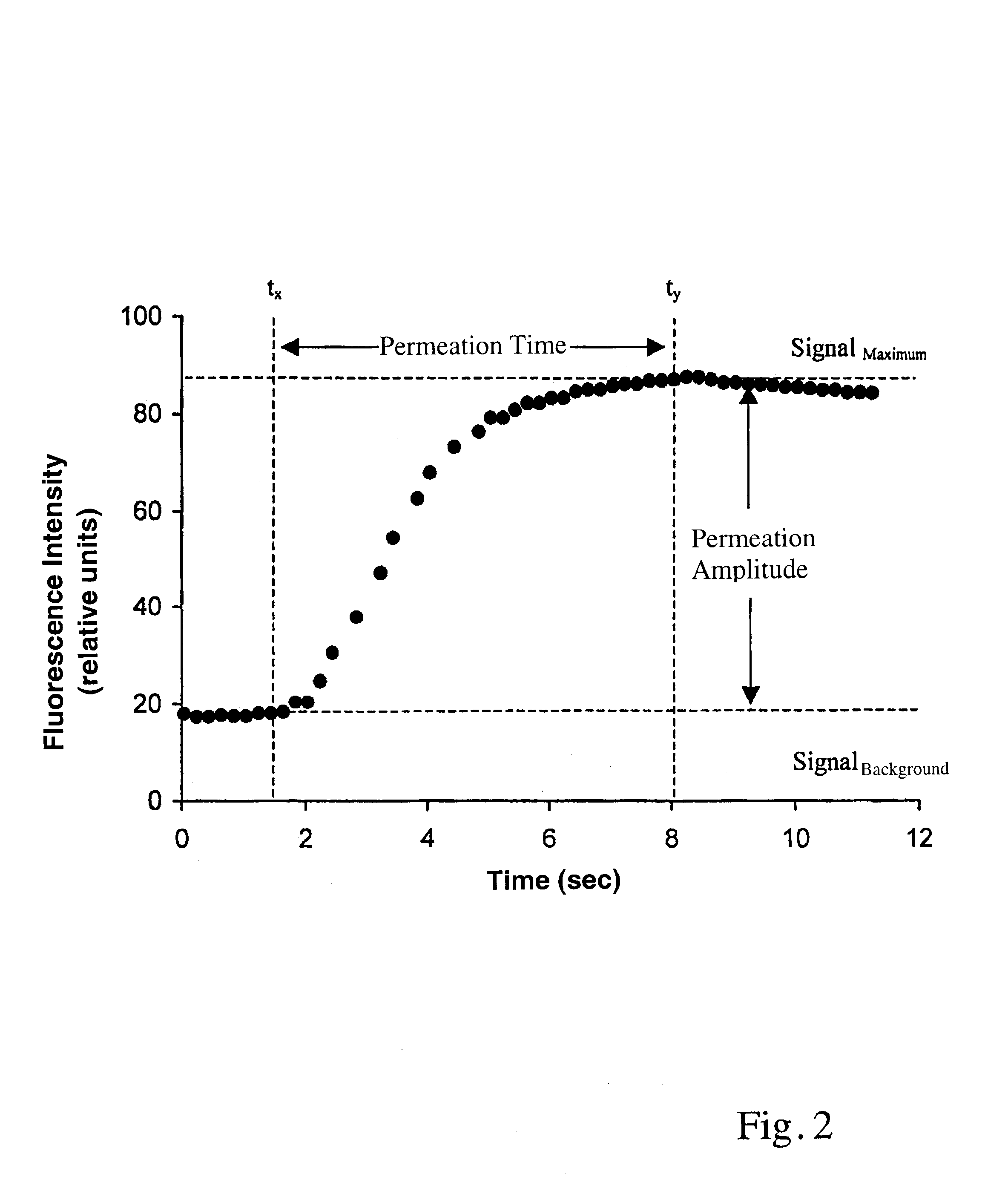
In a method, a device and a computer program for determining the blood flow in a tissue or organ region, the fluorescence intensity of an exogenous chromophore is measured as a function of time in the tissue region to be examined and from this the permeation rate of the chromophore is calculated, from which the blood flow can be derived.

Description

BACKGROUND OF THE INVENTIONThe present invention relates to a method, a device and a computer program for determining the blood flow in a tissue or organ region.A method for determining the depth of burns in tissue is known from U.S. Pat. No. 5,074,306, in which the fluorescence of the exogenous chromophore indocyanine green is detected and the depth of burn wounds is determined on the basis of its distribution in the tissue.In “Annals of Plastic Surgery”. 1999, Vol. 42, pp. 266-274 a device and a method for measuring the blood circulation through transferred skin flaps is described, wherein after applying indocyanine green the fluorescence stimulated by irradiation with a pulsed laser array is detected with a CCD camera.In the case of the known methods and known devices only the relative fluorescence distribution of the chromophore indocyanine green in the tissue is examined qualitatively to establish the blood circulation through the tissue. The known methods are not suitable for ...

Claims


Application Information

Patent Timeline
IPC IPC(8): A61B5/0275A61B5/026A61B5/00
CPCA61B5/0059A61B5/0275A61B5/0261
Inventor PFEIFFER, ULRICH J.BURGER, THORSTENBECKER, ANDREAS
Owner RENEW GRP
Learn More