Method and apparatus for evaluating medical examination images

a medical examination and image technology, applied in the field of medical examination image evaluation methods and apparatuses, can solve the problems of not 100% reliable, easy to occur, and images with abnormalities are overlooked, and achieve the effect of enhancing the dependability of examination image evaluation

Inactive Publication Date: 2005-03-08
SIEMENS HEALTHCARE GMBH
View PDF4 Cites 63 Cited by
  • Summary
  • Abstract
  • Description
  • Claims
  • Application Information

AI Technical Summary

Benefits of technology

An object of the present invention is to provide a medical image evaluation method and apparatus which avo

Problems solved by technology

This method, however, is not 100% reliable, so that all images must be monitored by a physician.
Due to the large number of images, it can easily occur that images having abnormalities are overlooked, since the individual images are viewed for only a very brief time.
Since t

Method used


Image

  • Method and apparatus for evaluating medical examination images
  • Method and apparatus for evaluating medical examination images

Examples


Embodiment Construction

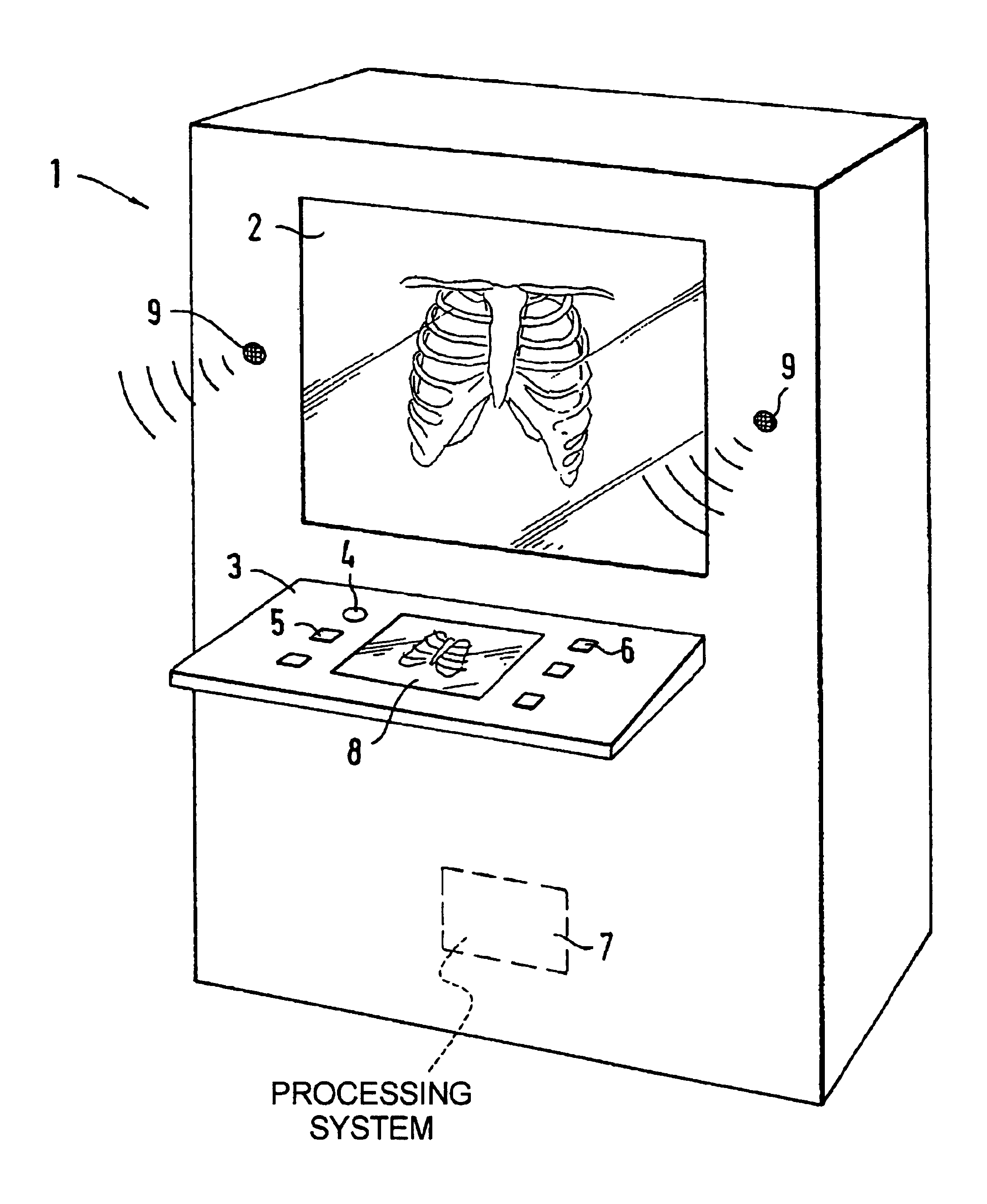
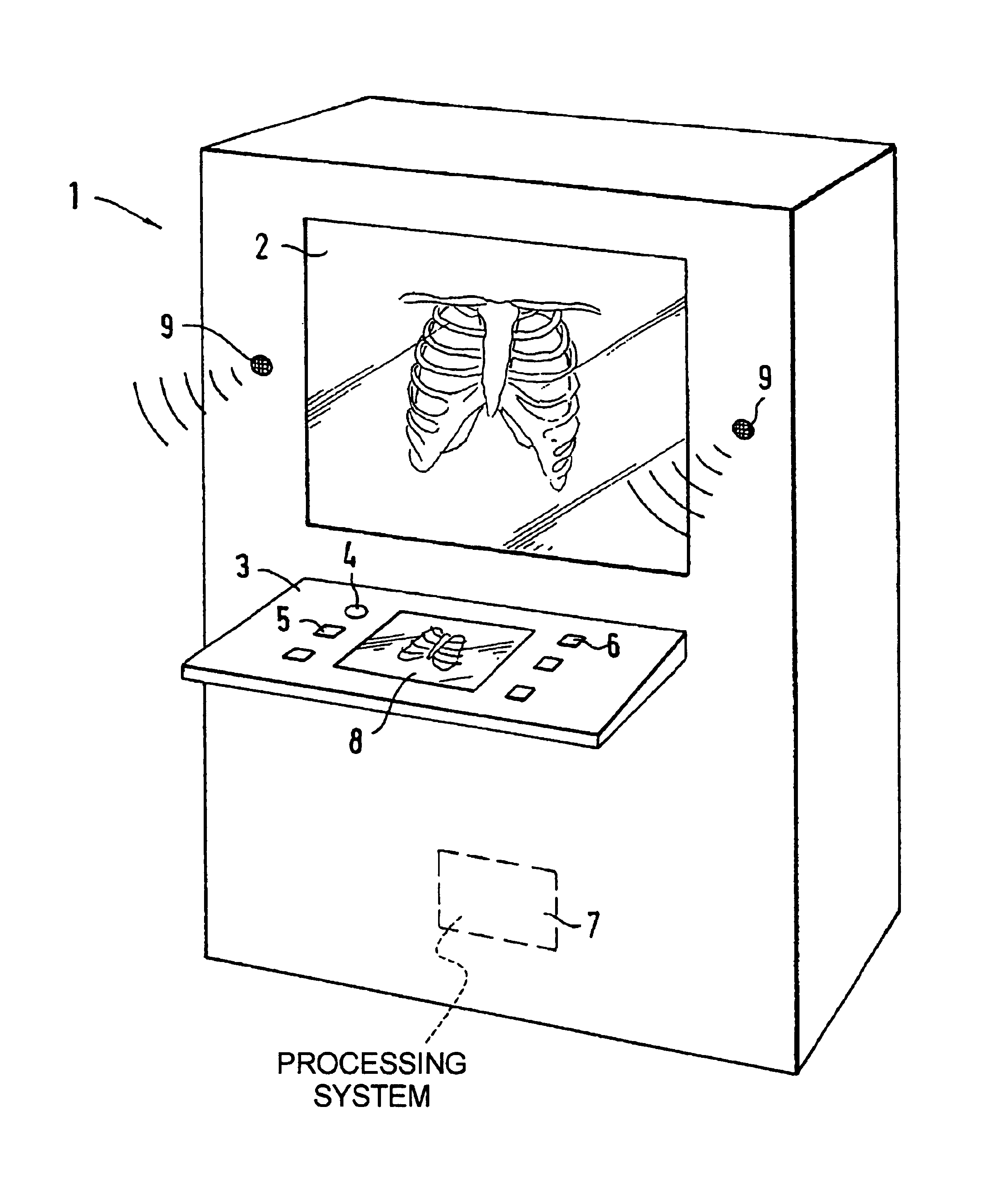
Medical examination images, for example x-ray images, can be presented at a viewing device 2 of the apparatus 1. A greater number of such images can be placed into the apparatus 1 and can be automatically sequentially presented. Each image is presented for a specific, relatively short time in order to be viewed by the physician. After the time has elapsed, the next image is automatically presented. Various presentation parameters can be set at a control panel 3 via a rotary knob 4 and input keys 5. Further input keys 6 are arranged at the control panel 3 that enable a marking of images with peculiarities in order to subsequently study these x-ray images exactly. The x-ray images are digitalized in the apparatus, i.e. are converted into computer-readable image data. To that end, the apparatus 1 contains a data processing device 7 (shown with broken lines) which is a high-performance computer. The data processing device 7 has software available with an evaluation algorithm for detecti...


PUM


Abstract

In a method and to an apparatus for evaluating a sequence of medical examination images successively presented at a viewing device using a data processing device operating on software having an evaluation algorithm for determining image regions exhibiting abnormalities, an acoustic signal is emitted by the dependent on the determination result, and/or the presentation time of an image on a monitor is influenced dependent thereon.

Description

BACKGROUND OF THE INVENTION1. Field of the InventionThe present invention is directed to a method and an apparatus for evaluating a sequence of medical examination images successively presented at a viewing device by means of a data processing device running on software having an evaluation algorithm for determining image regions exhibiting abnormalities.2. Description of the Prior ArtTechnical progress in medical technology has lead to a great increase of examination results in image form. There is a tendency to implement series examinations at a large number of patients, with a few images, for example, four examination images, being respectively for each patient. This is the case implemented, for example, in mammography and in lung examinations for detecting tuberculosis. By contrast, there are examination methods wherein tens of thousands of examination images from a single patient arise. Examples of this are tomography, MR, angiography and spiral CT. In all of these examination ...

Claims


Application Information

Patent Timeline
IPC IPC(8): A61B6/00G06T7/00
CPCA61B6/00A61B6/461G06T7/0012G06T2207/30068G06T2207/30061
Inventor BETZ, ROLAND
Owner SIEMENS HEALTHCARE GMBH
Why Eureka
Try Eureka