Method and apparatus for noninvasive intraductal fluid diagnostic screen

a non-invasive, intra-uterine fluid technology, applied in the field of non-invasive intra-uterine fluid diagnostic screen, can solve the problems of difficult detection of difficulty in identifying malignant and benign breast cancer

Inactive Publication Date: 2005-04-05
HHC PREFERRED
View PDF89 Cites 32 Cited by
  • Summary
  • Abstract
  • Description
  • Claims
  • Application Information

AI Technical Summary

Problems solved by technology

All too often, breast cancer is discovered at a stage that is too far advanced, when therapeutic options and survival rates are severely limited.
In addition to the problem of early detection, there remain serious problems in distinguishing between malignant and benign breast disease, in staging known breast cancers, and in differentiating between different types of breast cancers (e.g. estrogen dependent versus non-estrogen dependent tumors).
Previously, many cancer markers could only be detected or measured using conventional biochemical assay methods, which generally require large test samples and are therefore unsuitable in most clinical applications.
For the purpose of breast cancer evaluation, the use of conventional or needle biopsy samples for cancer marker assays is often undesirable, because a primary goal of such assays is to detect the cancer before it progresses to a palpable or mammographically detectable tumor stage.
Prior to this stage, biopsies are generally contraindicated, making early screening and low risk monitoring procedures employing such samples untenable.
Efforts to utilize serum samples for breast cancer marker assays have met with limited success.
The targeted markers are either not detectable in serum, or telltale changes in the levels or activity of the markers cannot be monitored in serum.
In addition, the presence of breast cancer markers in serum may occur at the time of micro-metastasis, making serum assays less useful for detecting pre-metastatic disease.
Although the evaluation of mammary fluid has been shown to be a useful method for screening nonpalpable breast cancer in women who experience spontaneous nipple discharge, the rarity of this condition renders the methods of Inaji et al, inapplicable to the majority of women who are candidates for early breast cancer screening.
It is likely that other antibodies used to assay other cancer markers may exhibit even lower sensitivity than the anti-CEA antibodies used by Inaji and coworkers, and may therefore not be adaptable or sensitive enough to be employed even in dry chemical immunoassays of small samples of spontaneously discharged mammary fluid.

Method used


Image

  • Method and apparatus for noninvasive intraductal fluid diagnostic screen
  • Method and apparatus for noninvasive intraductal fluid diagnostic screen
  • Method and apparatus for noninvasive intraductal fluid diagnostic screen

Examples


Embodiment Construction

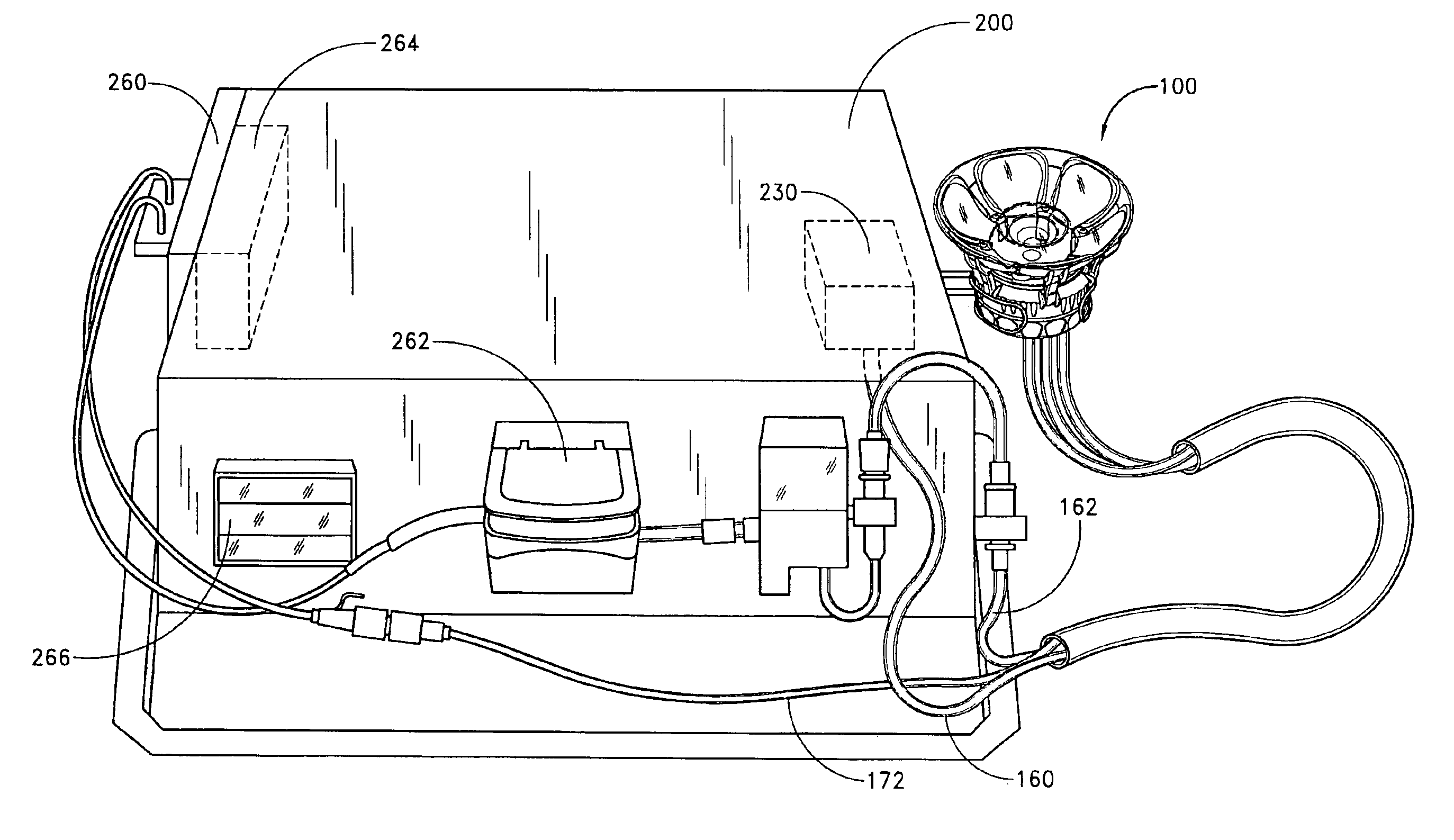
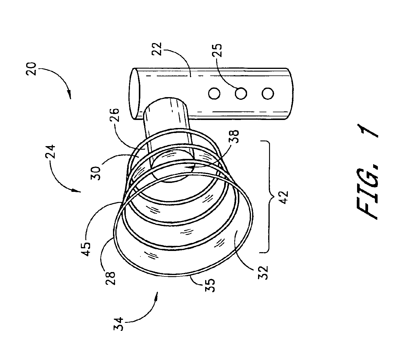
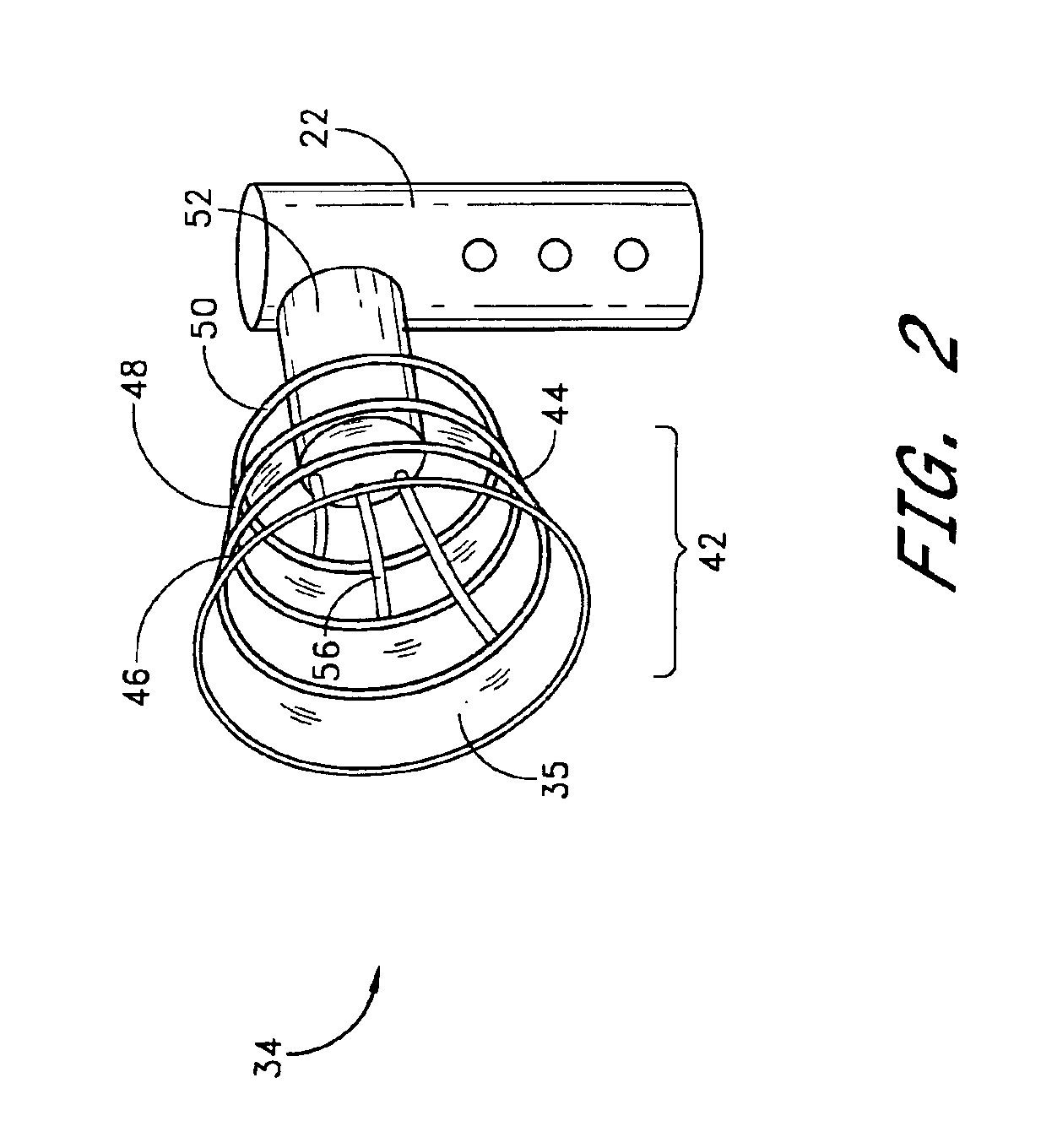
Referring to FIG. 1, there is illustrated a schematic representation of a portable, self-contained intraductal fluid aspiration device 20 in accordance with one aspect of the present invention. The aspiration device 20 includes a housing 22, for containing various controls and functional components of the device 20. One or more controls and / or indicators 25 may be provided on the housing, for controlling various aspects of the device such as suction, compression, and other features (e.g., heat, ultrasound) which may be included depending upon the intended functionality of the aspiration device 20. The housing 22 may be formed by extrusion, injection molding or other well known techniques from a suitable biocompatible material such as high density polyethylene, nylon, polyethylene terephthalate, or others well known in the art. The housing is preferably formed in an ergonomic configuration, to comfortably facilitate grasping in one hand during use.

The housing 22 is provided with a pa...


PUM


Abstract

Disclosed is a method and apparatus for accomplishing a noninvasive screen for breast disease markers, including breast cancer markers and cytologically abnormal cells. Intraductal fluid is noninvasively aspirated using compression, heating and suction cycles. The removed sample is thereafter assayed for the presence of cytologically abnormal cells and / or one or more breast disease markers. In addition to the diagnostic applications of the disclosed method and apparatus, a therapeutic application is also contemplated wherein intraductal fluids are noninvasively aspirated using compression, heating and suction cycles to remove toxic buildups within the fluid.

Description

BACKGROUND OF THE INVENTIONBreast cancer is by far the most common form of cancer in women, and is the second leading cause of cancer death in humans. Despite many recent advances in diagnosing and treating breast cancer, the prevalence of this disease has been steadily rising at a rate of about 1% per year since 1940. Today, the likelihood that a woman living in North America will develop breast cancer during her lifetime is one in eight. The current widespread use of mammography has resulted in improved detection of breast cancer. Nonetheless, the death rate due to breast cancer has remained unchanged at about 27 deaths per 100,000 women. All too often, breast cancer is discovered at a stage that is too far advanced, when therapeutic options and survival rates are severely limited. Accordingly, more sensitive and reliable methods are needed to detect small (less than 2 cm diameter), early stage, in situ carcinomas of the breast. Such methods should significantly improve breast can...

Claims


Application Information

Patent Timeline
Patent Type & Authority Patents(United States)
IPC IPC(8): A61B10/00A61M1/06A61B10/02
CPCA61B5/6834A61B10/0041A61B10/0045A61M1/066A61M1/064A61M1/06A61M2205/3653A61B10/0283A61M1/0697A61M1/0693
Inventor MORTON, KEVIN B.BARE, REX O.SMITH, JEFFREY C.PAYNE, TIMOTHY J.GLEASON, PAUL
Owner HHC PREFERRED
Why Eureka
Try Eureka