
Biopsy device

a biopsy and device technology, applied in medical science, vaccination/ovulation diagnostics, therapy, etc., can solve the problems of reducing the likelihood of an adequate yield, limiting the therapeutic options and survival rates, and only finding breast cancer imaging techniques

Inactive Publication Date: 2005-05-31
LACHOWICZ THEODORE COLLATERAL AGENT
View PDF8 Cites 238 Cited by
  • Summary
  • Abstract
  • Description
  • Claims
  • Application Information

AI Technical Summary

Benefits of technology

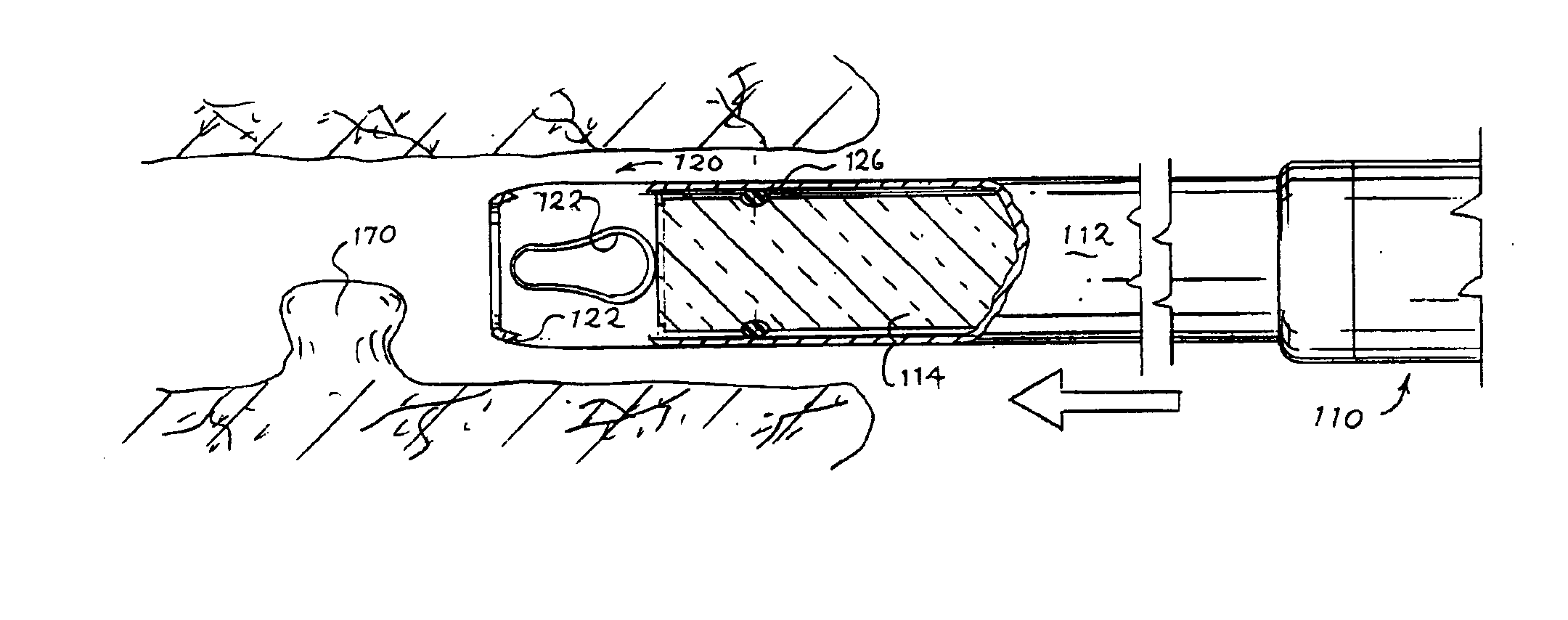
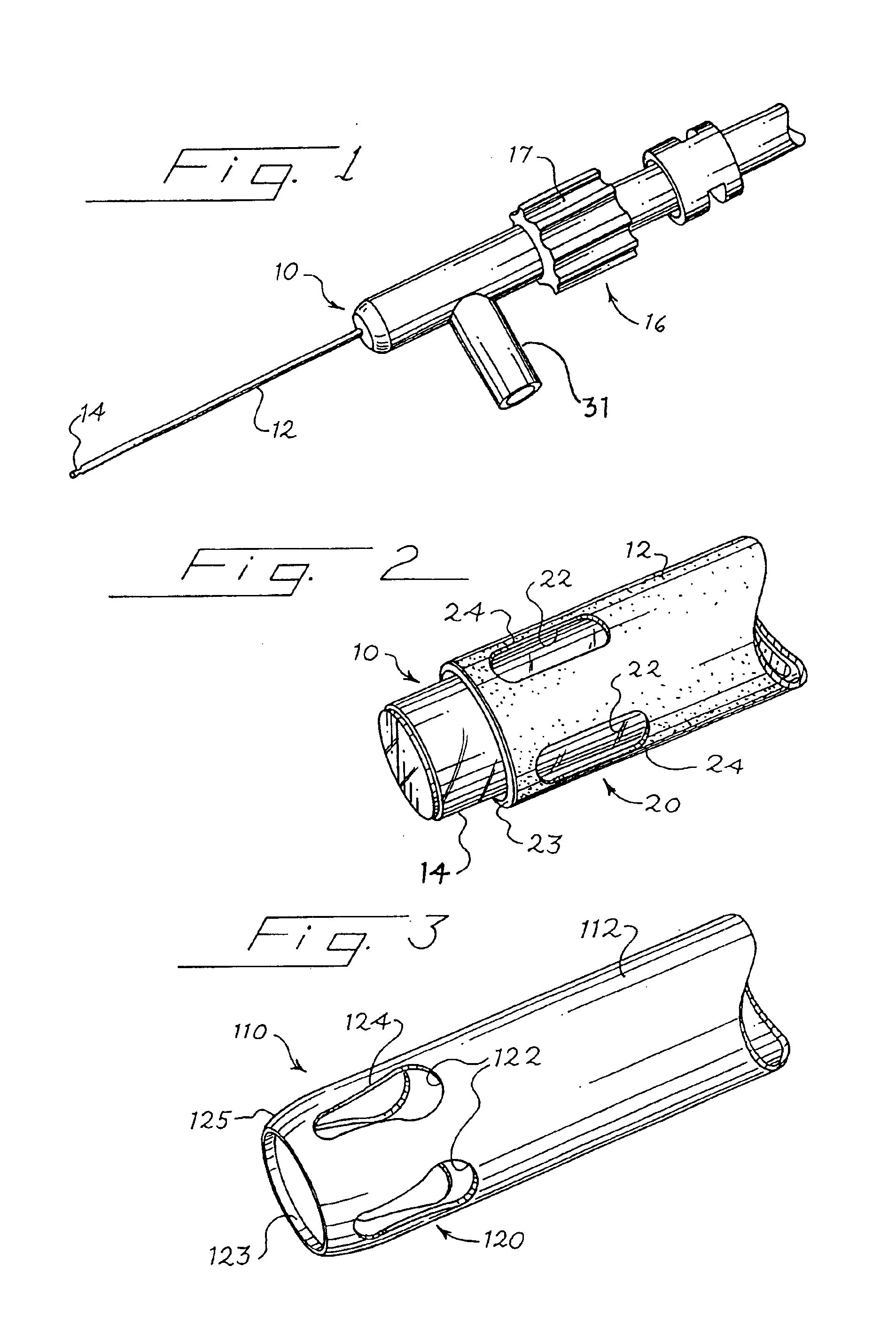
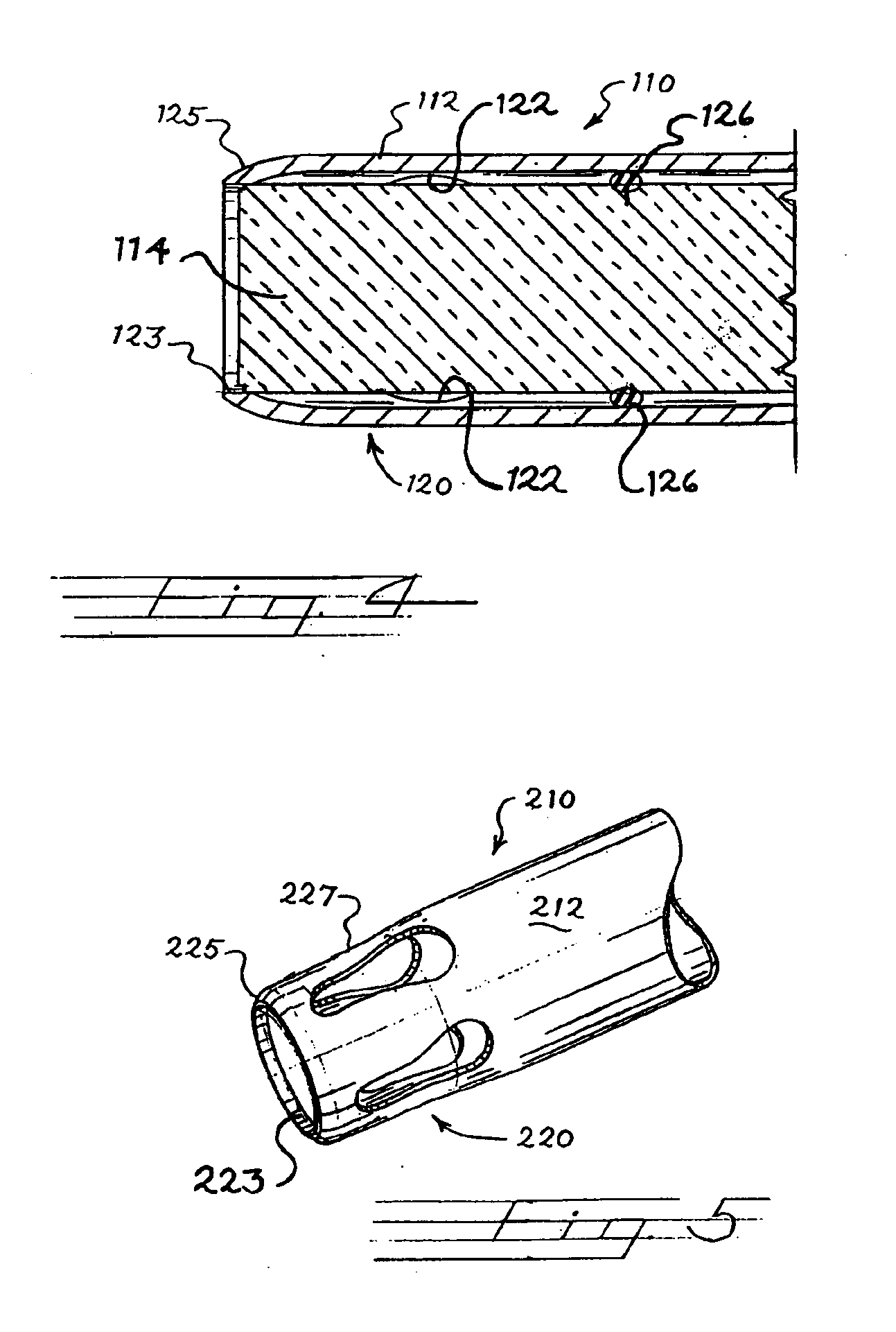
[0008]The endoscope extends through the sheath and may be extended beyond the distal end of the sheath. Preferably during insertion of the device into a mammary duct, the endoscope is positioned such that the distal end of the endoscope is substantially co-terminus with the distal end of the sheath. As such, the endoscope and sheath will have few, if any, sharp edges on the leading edge of the device and may therefore act as a mammary duct dilator and obturator with minimal risk of injury to the mammary duct. Such precise alignment of the endoscope further permits accurate location of the working end of the device within the mammary duct. The endoscope can also be extended beyond the distal end of the sheath. In order to further lessen the likelihood of ductal injury during the insertion of the device, the distal end portion terminates in an atraumatic tip including a circumferential taper, a circumferential bevel or both.
[0010]A brushing biopsy is performed with the present invention by rotating the device within the mammary duct. Prior to insertion of the present invention into the mammary duct, it is preferred that the endoscope is positioned to be substantially co-terminus with the distal end of the sheath. A physician or other medical person views the mammary duct as the device of the present invention is inserted using a viewing device operatively connected to the endoscope, such as a manual lens or a video display. When the distal end portion of the sheath is situated in the desired region of the mammary duct, the physician or other medical person retracts the endoscope to expose the plurality of apertures on the distal end portion of the sheath, and rotates the sheath. In so doing, the edges of the apertures, and in some embodiments a roughened surface of the sheath, act to brush the intraductal epithelium and loosen cytologic material suitable for analysis. Preferably, the endoscope is retracted such that the apertures in the sheath are fully exposed and the physician is able to view the mammary duct to target accurately the desired area or region to be brushed.

Problems solved by technology

One problem with mammography is that such an imaging technique can only find breast cancer once it has taken form.
All too often, breast cancer is discovered at a stage that is too far advanced, when therapeutic options and survival rates are severely limited.
The soft nature of the bristles and the absence of adequate radial force directing the bristles into the epithelium reduce the likelihood of an adequate yield.
Such brush biopsy devices also offer no papilloma or carcinoma biopsy capability because they cannot effectively excise a tissue sample from the area of stricture.

Method used


Image

  • Biopsy device
  • Biopsy device
  • Biopsy device

Examples


Embodiment Construction

[0030]The invention disclosed herein is susceptible of being embodied in many different forms. Shown in the drawings and described herein below in detail are preferred embodiments of the invention. It is to be understood, however, that the present disclosure is an exemplification of the principles of the invention and does not limit the invention to the illustrated embodiments.

[0031]Referring to FIGS. 1 and 2, biopsy device 10 comprises a sheath 12 rotatable about a longitudinal axis and an endoscope 14 extending through the sheath 12. An adjustment mechanism 16 is also operatively connected to the endoscope 14 to longitudinally extend and retract the endoscope 14. The endoscope 14 may act as a dilator as well as an obturator. In a preferred embodiment, the endoscope 14 is extended and retrapted within the sheath 12 by a rotatable positioning hub 17. It is also preferred that the rotatable positioning hub 17 can be locked in place so as to fix the position of endoscope 14 relative t...


PUM


Abstract

A biopsy device suitable for collecting cells from a mammary duct is provided. The device is suitable for conducting brushing biopsy and tissue excision procedures. The device comprises a sheath that is rotatable about a longitudinal axis. The device further comprises an endoscope disposed within and extending through the sheath. An adjustment mechanism is also operatively connected to the endoscope to longitudinally extend and retract the endoscope within the sheath.

Description

TECHNICAL FIELD OF THE INVENTION[0001]The invention relates to a device for obtaining cytologic samples. More specifically, the invention relates to a biopsy device utilizing a viewing component.BACKGROUND OF THE INVENTION[0002]Breast cancer is one of the health threats most feared by women, and is the most common form of cancer in women. A key to treatment is early detection. For example, an annual mammogram is a method that has been used in hopes of early detection of breast cancer. One problem with mammography is that such an imaging technique can only find breast cancer once it has taken form. All too often, breast cancer is discovered at a stage that is too far advanced, when therapeutic options and survival rates are severely limited. As such, more sensitive and reliable methods and devices are needed to detect cancerous, pre-cancerous, and other cancer markers of the breast at an early stage. Such methods and devices could significantly improve breast cancer survival. While b...

Claims


Application Information

Patent Timeline
Patent Type & Authority Patents(United States)
IPC IPC(8): A61B10/00A61B19/00A61M1/00A61B10/02
CPCA61B10/0041A61B10/0266A61B10/0275A61B10/0045A61B2217/007A61B2019/5231A61B2217/005A61B2090/373
Inventor KERMODE, JAMESMUELLER, JR., RICHARD L.
Owner LACHOWICZ THEODORE COLLATERAL AGENT
Learn More