Historical comparison of breast tissue by image processing

a breast tissue and image processing technology, applied in the field of three-dimensional, ultrasonic mammography, can solve the problems of difficult to determine whether the same lesion is comparable in earlier and later images, the comparison and even interpretation of images is complicated, and the breast may move or be slightly deformed

Inactive Publication Date: 2005-06-21
LITTON SYST INC +1
View PDF15 Cites 131 Cited by
  • Summary
  • Abstract
  • Description
  • Claims
  • Application Information

AI Technical Summary

Benefits of technology

[0018]FIG. 9 is a symbolic diagram of an optical correlator, optionally used in accordance with a variation of the invention

Problems solved by technology

Comparison and even interpretation of images is complicated in part because the region of interest is a three-dimensional volume, while the image is a projection onto a two-dimensional plane or planes.
Additional difficulties result because the breast may move slightly or be slightly deformed between the earlier and later images.
Ev

Method used


Image

  • Historical comparison of breast tissue by image processing
  • Historical comparison of breast tissue by image processing
  • Historical comparison of breast tissue by image processing

Examples


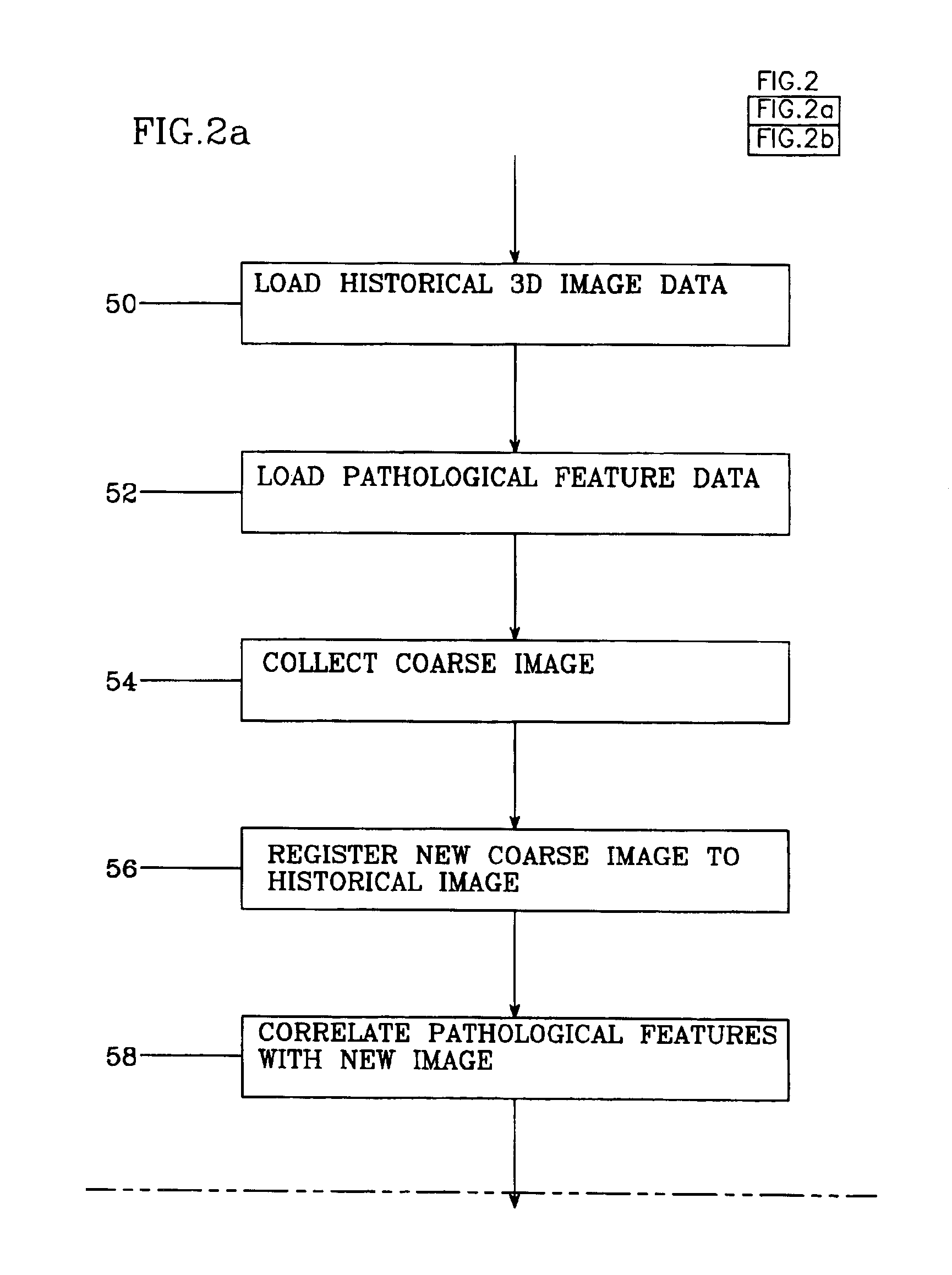
Embodiment Construction

Overview of Apparatus

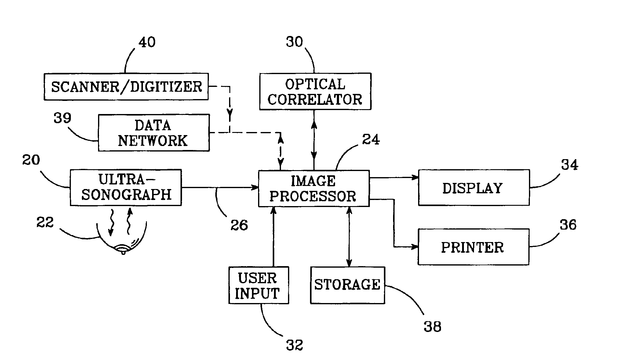
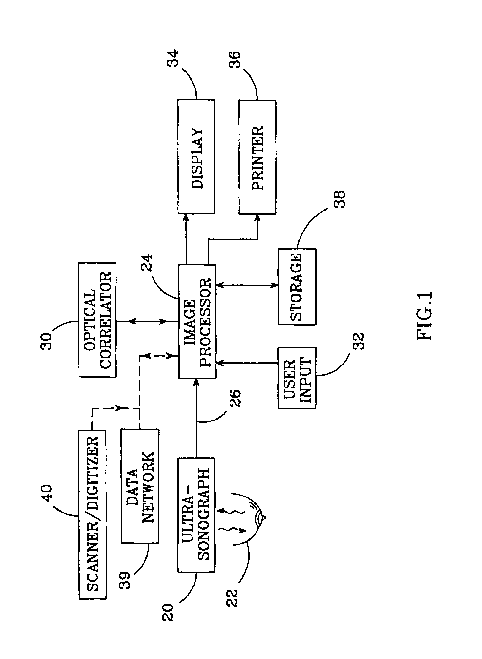
[0019]FIG. 1 shows an apparatus in accordance with the invention and suitable for practicing its method. An ultrasonographic imaging system 20 (or its equivalent) images a patient's breast 22. The imaging system 20 provides digital image data to an image processor 24 via an input channel 26. An optical correlator 30 is preferably interfaced with and controlled by the image processor 24 to provide high-speed image processing (correlations) of pre-processed image data. A user input device 32 (typically a keyboard and / or graphical pointing device such as a mouse) is interfaced to the image processor 24 to allow user control of the image processor 24. Graphic output is displayed by the image processor 24 on a display 34, which is preferably a color-capable video display. A printer36 is preferably also interfaced with image processor 24 to produce “hard copy” printouts which record the imagery, most preferably with multi-color, high resolution graphics. A storage dev...


PUM


Abstract

An image processing system and method visually documents and displays changes between historical and later mammographic images, preferably in three dimensions. A composite image is created which visually emphasizes temporal differences between the historical and later images. Preferably three-dimensional, digitized images, displayable in various projections, are stored for archival purposes on computer readable media. An image processor preferably exploits an optical correlator to register the historical and later images accurately and provide correlation values as temporal scalars of the differences. The registered images are then compared, voxel-by-voxel, to detect temporal differences. The composite image is displayed with synthetic colors or other visual clues to emphasize apparent changes (for example, tumor growth or shrinkage).

Description

BACKGROUND OF THE INVENTION[0001]1. Field of the Invention[0002]The invention relates generally to ultrasonic and radiographic medical imaging, and particularly to three-dimensional, ultrasonic mammography for the purpose of detecting and visualizing breast lesions.[0003]2. Description of the Related Art[0004]The standard of care for managing breast disease includes (in order) detection, diagnostic work-up, definitive diagnosis, treatment, adjuvant therapy and follow-up. In a large number of cases, the health care provider will not consider a biopsy justified and rather will opt to follow the situation with examinations every three to six months. In other cases the health care provider will typically also want to follow the progress of the lesion after adjuvant therapies are administered. This implies repeated imaging of the region of tissue containing the lesion and comparison of the earlier and later images, to detect and characterize any changes in the lesion.[0005]Various imagin...

Claims


Application Information

Patent Timeline
IPC IPC(8): G06K9/00G06K9/32G06T7/00
CPCG06T7/0012G06T7/0026G06T2207/30068G06T7/32A61B5/4848A61B6/502
Inventor CARROTT, DAVID T.BURKE, THOMAS M.
Owner LITTON SYST INC
Why Eureka
Try Eureka