Integrated multi-modality imaging system

a multi-modality imaging and imaging system technology, applied in the field of imaging systems, can solve the problems of not being able to accurately determine not being able to provide tissue imaging indicative of the actual location of the probe, and being unable to provide the type of information and feedback that is needed for providing the type of information and feedback

Inactive Publication Date: 2005-08-02
GENERAL ELECTRIC CO
View PDF4 Cites 58 Cited by
  • Summary
  • Abstract
  • Description
  • Claims
  • Application Information

AI Technical Summary

Benefits of technology

[0006]The present invention provides an integrated imaging system designed to respond to these needs. The technique may be applied as a combination of various different imaging modalities, but is particularly well suited to combining MRI systems with x-ray-based systems, such as digital x-ray fluoroscopy systems. The systems are combined in a complimentary and cooperative manner, such that real-time images may be produced of soft tissues through use of MRI imaging sequences, while images of more dense or contrasting tissues and objects may be produced through the x-ray system. The systems may be physically combined by positioning a specially-adapted support structure for a digital x-ray apparatus in an MRI system. Separate images may be produced by the two systems, with the desired anatomy of soft tissues being projected on a screen associated with the MRI system, while the x-ray image is displayed on a separate screen. Alternatively, the system may be adapted to register and combine the images to provide real-time feedback of all of the structures of interest, thereby making use of the strength of the combined modalities in the resulting imaging. In addition to real-time imaging, the system may be employed to produce images which are registered or associated with one another, with the still images being available for viewing, storage, transmission, and so forth.

Problems solved by technology

These contrast agents, however, do not necessarily provide the particular tissue identification desired, may not be retained for the time and in the locations desired for the entire procedure, and may cause complications for certain patients.
For example, during catheterization, angioplasty, and similar procedures, MRI systems may permit a surgeon to identify soft tissues through which a probe is inserted, but are not necessarily well suited to imaging tissues indicative of the actual location of the probe.
Because surgical interventions happen in real time, currently available technologies for separate modality imaging are simply ill suited to providing this type of information and feedback.

Method used


Image

  • Integrated multi-modality imaging system
  • Integrated multi-modality imaging system
  • Integrated multi-modality imaging system

Examples


Embodiment Construction

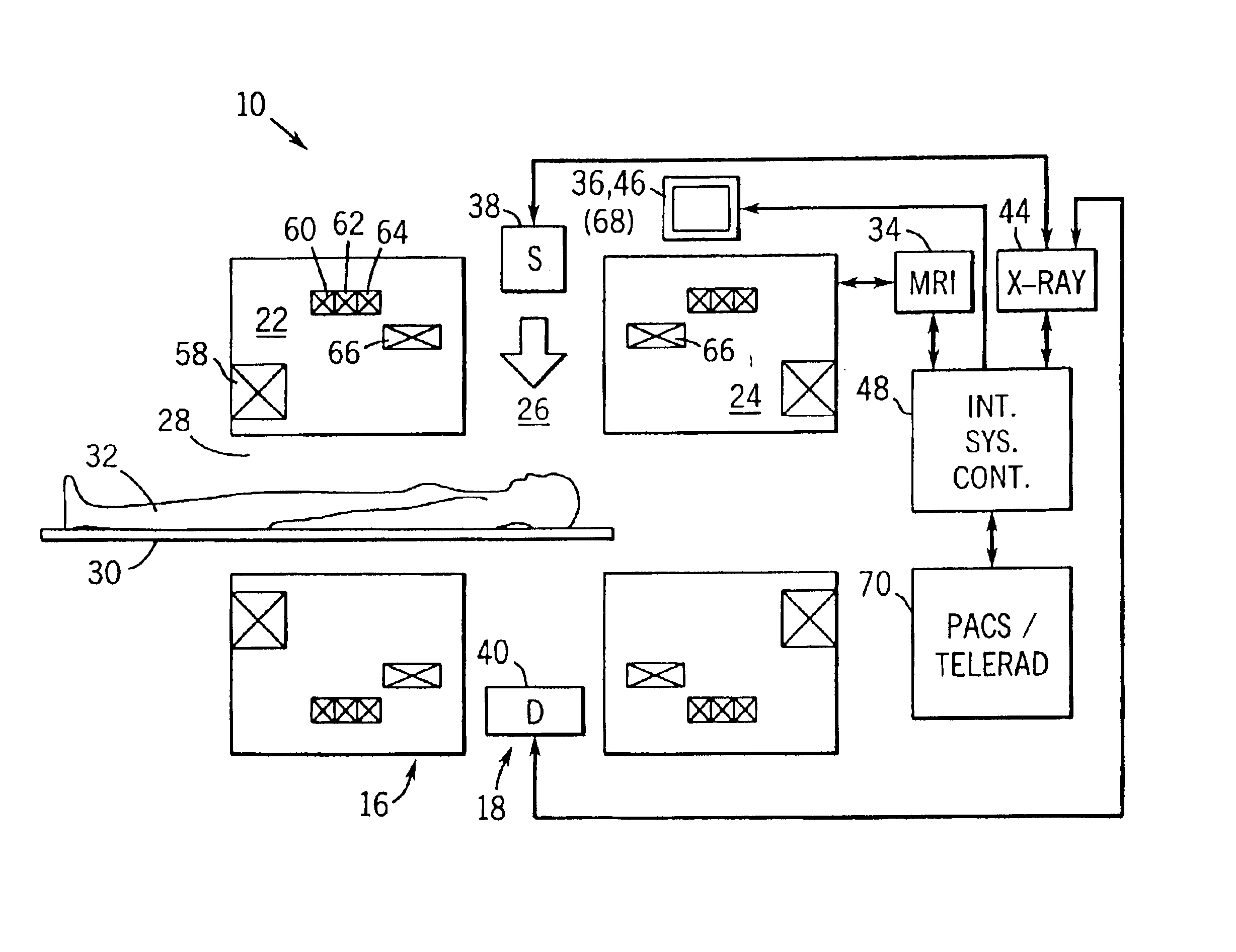
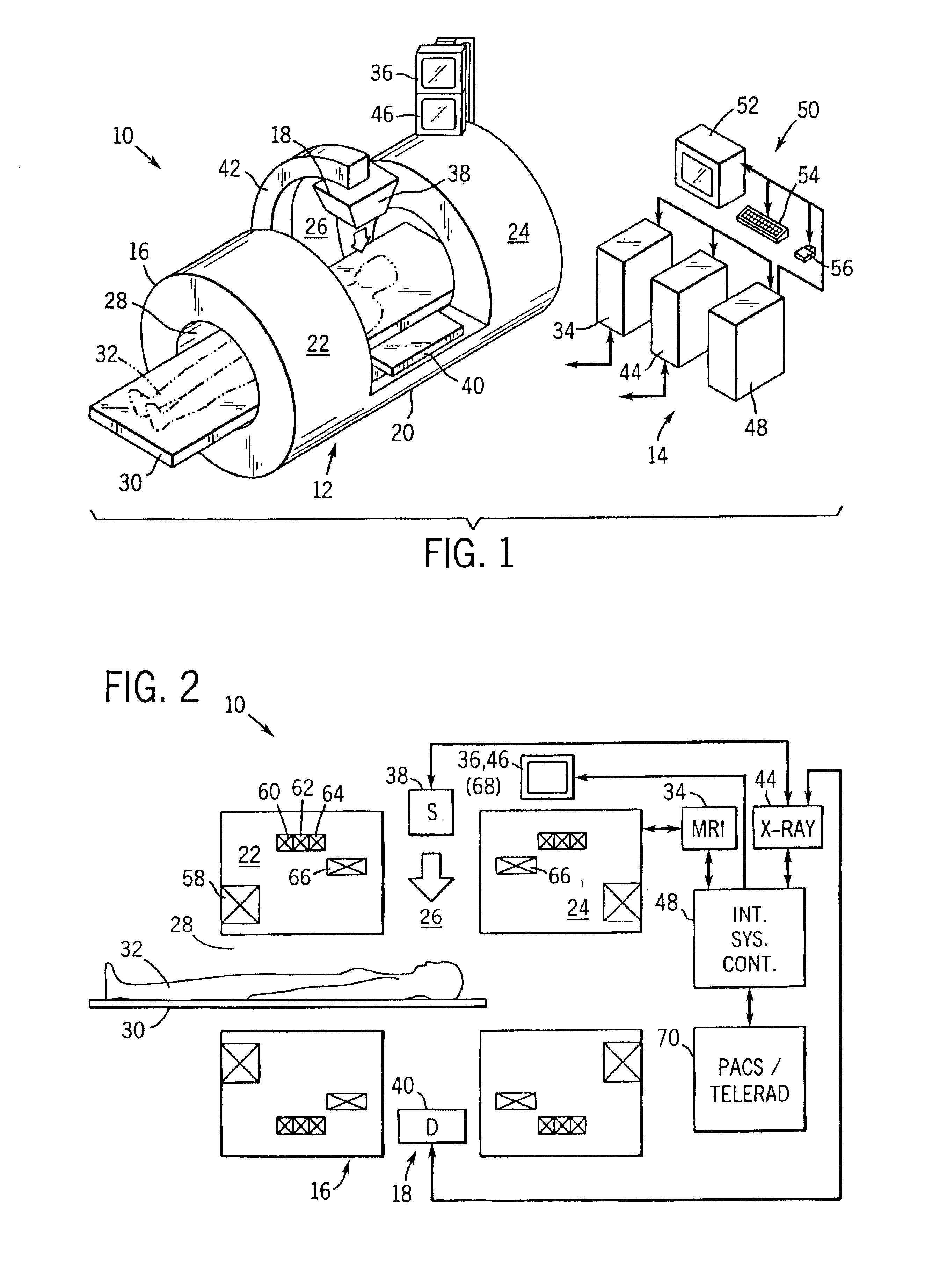
[0011]Turning now to the drawings, and referring first to FIG. 1, an integrated, multi-modality imaging system 10 is illustrated as including a data acquisition station 12 and a data processing and control station 14. In the illustrated embodiment, the imaging system includes components of magnetic resonance imaging (MRI) and x-ray modalities. Specifically, the system includes a split magnet MRI system 16 and a digital x-ray system 18 configured to generate images during a medical procedure to provide feedback to a medical diagnostic or surgical team. It should be noted, however, that while the MRI and x-ray modalities described herein are combined by way of example, various other modalities may be combined in similar manners to draw upon the strengths of the particular imaging modalities involved in viewing specific tissues, surgical devices, anatomical features and physiological functions.

[0012]In the arrangement illustrated in FIG. 1, MRI system 16 includes a coil housing 20 whic...


PUM


Abstract

An integrated, multi-modality imaging technique is disclosed. The embodiment described combines a split-magnet MRI system with a digital x-ray system. The two systems are employed together to generate images of a subject in accordance with their individual physics and imaging characteristics. The images may be displaced in real time, such as during a surgical intervention. The images may be registered with one another and combined to form a composite image in which tissues or objects difficult to image in one modality are visible. By appropriately selecting the position of an x-ray source and detector, and by programming a desired corresponding slice for MRI imaging, useful combined images may be obtained and displayed.

Description

CROSS-REFERENCE TO RELATED APPLICATIONS[0001]This application is a continuation of U.S. application Ser. No. 09 / 268,401, filed Mar. 15, 1999, now issued as U.S. Pat. No. 6,591,127.FIELD OF THE INVENTION[0002]The present invention relates generally to the field of imaging systems such as those used in medical diagnostics. More particularly, the invention relates to an integrated system that makes use of different modalities in a complementary fashion to permit feedback to surgeons and other medical professionals of physical conditions of a subject, particularly during interventionary procedures.BACKGROUND OF THE INVENTION[0003]A wide variety of imaging systems have been developed and are presently in use in the medical field. The systems may be generally categorized in a series of “modalities,” with each modality being characterized by its particular physics, control, utility, and so forth. For example, magnetic resonance imaging (MRI) systems are commonly employed for producing imag...

Claims


Application Information

Patent Timeline
Patent Type & Authority Patents(United States)
IPC IPC(8): A61B5/055A61B6/00A61B5/05
CPCA61B6/00A61B6/5247G01R33/4812A61B6/4417
Inventor MCKINNON, GRAEME C.
Owner GENERAL ELECTRIC CO
Why Eureka
Try Eureka