Ultrasonic imaging apparatus and method

a technology of ultrasonic imaging and sub-aperture, which is applied in the field of ultrasonic imaging apparatus and methods, can solve the problem of not being able to completely unify the focal range of each and every receive sub-aperture, and achieve the effect of minimizing the difference in the delay time during beamforming

Inactive Publication Date: 2005-08-09
FUJIFILM HEALTHCARE CORP
View PDF7 Cites 22 Cited by
  • Summary
  • Abstract
  • Description
  • Claims
  • Application Information

AI Technical Summary

Benefits of technology

[0024]In this ultrasonic imaging method, the plurality of oscillators of the ultrasonic probe may be grouped such that the difference in delay time during the beamforming is minimized.

Problems solved by technology

However, since the number of elements in the entire group of apertures is limited, it is impossible to completely unify the focal ranges in each and every receive sub-aperture.

Method used


Image

  • Ultrasonic imaging apparatus and method
  • Ultrasonic imaging apparatus and method
  • Ultrasonic imaging apparatus and method

Examples


Embodiment Construction

[0037]The invention will be hereafter described by way of embodiments with reference made to the attached drawings, in which elements or parts having similar functions are referenced by similar numerals.

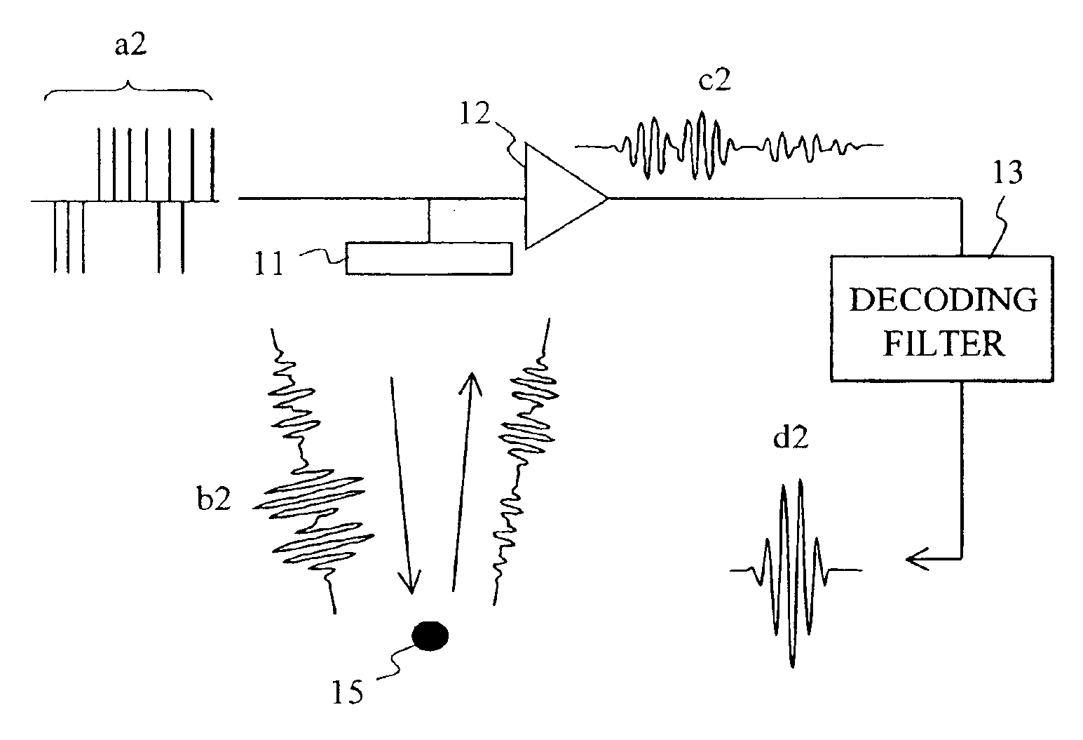
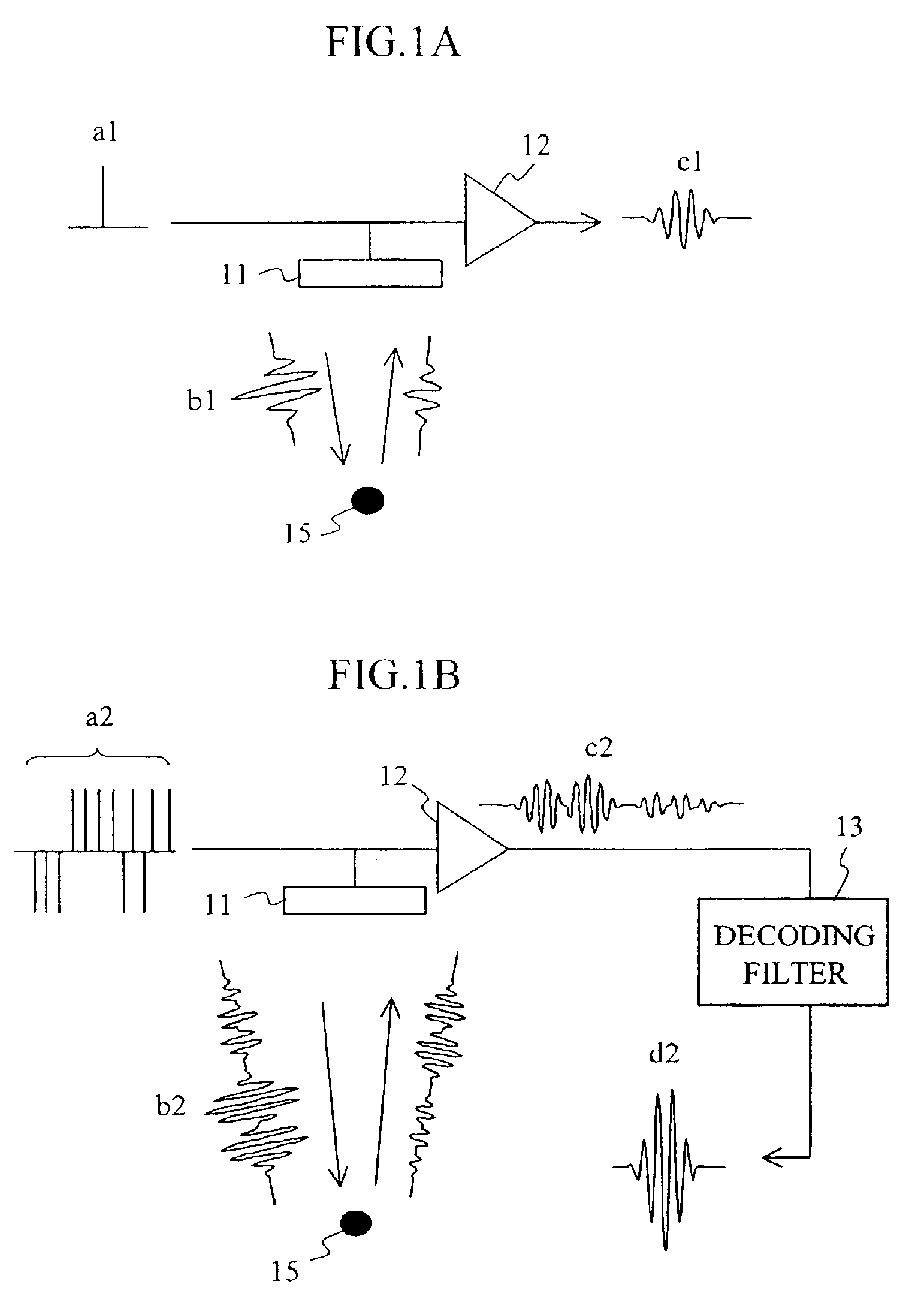
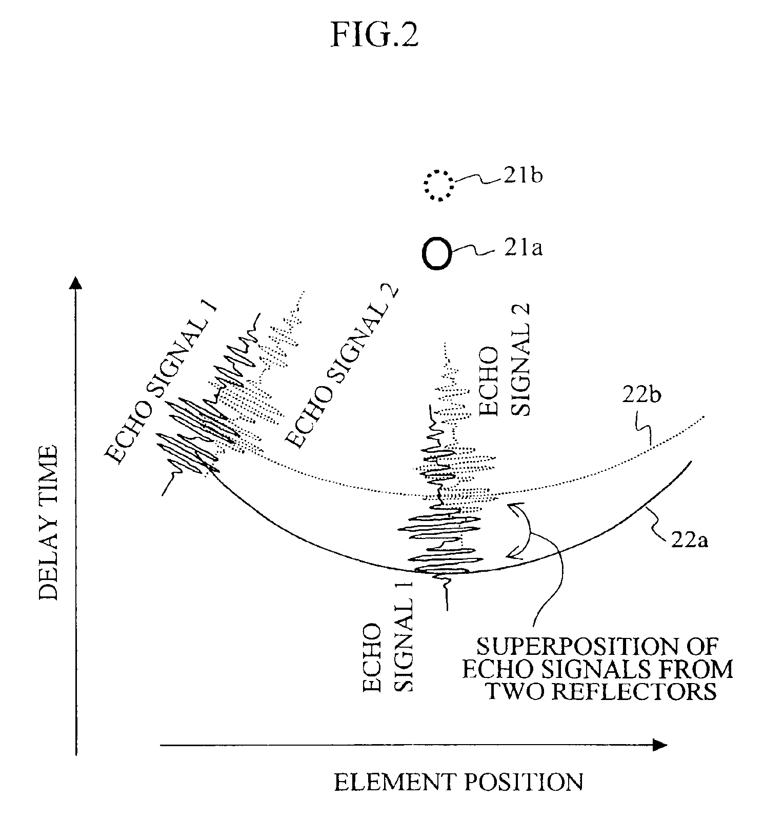
[0038]FIG. 3 shows a block diagram of an example of the ultrasonic imaging apparatus according to the invention. An ultrasonic probe 31 transmits a beam of ultrasonic waves to a subject (not shown) and receives an echo signal. A transmit signal (coded pulse signal) is read from a transmit waveform memory 32 and sent by the transmit beam former 33 via a group of transmission / reception switches 35 to the ultrasonic probe 31 under the control of a control system 34, with a delay time adapted to the transmit focusing point corresponding to the signal. The ultrasonic signal reflected or scattered inside the subject is returned to the ultrasonic probe 31. The ultrasonic probe 31 converts the ultrasonic signal into an electric signal. The electric signal is divided into receive sub-beam for...


PUM


Abstract

An ultrasonic imaging apparatus and method combines dynamically focused reception and coded transmission/reception technologies by inexpensive circuitry. A receive aperture is divided into smaller apertures. Correspondingly, a receive beam former is divided into receive sub-beam formers 36a to 36n by which a phase alignment and summing process is performed. Thereafter, coded signals are compressed in decoders 37a to 37n in a time axis direction. Output signals from the decoders are once again subjected to a phase alignment and summing process in a second beam former.

Description

BACKGROUND OF THE INVENTION[0001]1. Technical Field[0002]The present invention relates to ultrasonic imaging apparatus and methods for imaging internal organs of a sample body using ultrasound.[0003]2. Background Art[0004]Ultrasonic imaging apparatus, such as those used for medical diagnostical imaging, utilize ultrasonic pulse reflection to display a tomographic image of soft tissues of a living body or an image of blood flowing in a living body for observation on a monitor practically in real time. Ultrasonic imaging apparatus are considered highly safe, because they do not expose the subject to radiation as diagnostical imaging apparatus employing radiation. In addition, they are small and inexpensive. For these reasons, ultrasonic imaging apparatus are widely used in the field of medicine.[0005]An ultrasonic imaging apparatus produces images in the following way. An ultrasonic wave is transmitted to a subject from an ultrasonic probe made up of an array of piezoelectric elements...

Claims


Application Information

Patent Timeline
Patent Type & Authority Patents(United States)
IPC IPC(8): G01S15/89G01S15/00G01S7/52G10K11/34G10K11/00A61B8/00H04R3/00H04R17/00
CPCG01S7/52047G01S7/52093G01S15/8927G01S15/8959G10K11/341
Inventor AZUMA, TAKASHIUMEMURA, SHIN-ICHIROMIWA, YUICHI
Owner FUJIFILM HEALTHCARE CORP
Why Eureka
Try Eureka