Shape lockable apparatus and method for advancing an instrument through unsupported anatomy

a lockable, instrument technology, applied in the field of apparatus and methods for placing and advancing diagnostic or therapeutic instruments, can solve the problems of affecting the accuracy of the diagnosis, the risk of tissue capture or pinching, and the inability to advance the colonoscope as far as the cecum in up to one-sixth of all cases

Inactive Publication Date: 2005-09-13
USGI MEDICAL
View PDF78 Cites 250 Cited by
  • Summary
  • Abstract
  • Description
  • Claims
  • Application Information

AI Technical Summary

Benefits of technology

[0021]In view of the foregoing, it is an object of the present invention to provide apparatus and methods for facilitating placement of diagnostic or therapeutic instruments within easily distensible or unpredictably supported hollow body organs, such as the esophagus or colon.
[0022]It is a further object of the present invention to provide apparatus and methods that permit a diagnostic or therapeutic device to be advanced into a hollow body organ, and which facilitates passage of the device through tortuous anatomy without requiring straightening of organ passageways already traversed.
[0023]It also is an object of the present invention to provide apparatus and methods for facilitating placement of diagnostic or therapeutic instruments within easily distensible hollow body organs that include means for reducing the risk that tissue will become inadvertently pinched or caught as the diagnostic or therapeutic instrument is maneuvered through the hollow body organ.

Problems solved by technology

Problems regularly occur, however, when negotiating the colonoscope through the bends of the colon, such as at the sigmoid and left and right colic flexures.
These problems arise because the colon is soft and has unpredictable fixation points to the viscera of the abdomen, and it is easily distensible.
Consequently, the colonoscope cannot be advanced as far as the cecum in up to one-sixth of all cases.
Many patients find the operation of such previously-known devices unpleasant because the sigmoid portion of the colon is forced into an almost rectilinear shape by the guide tube.
Due to the stiffness of the guide tube, careless handling of the guide tube presents a risk of injury to the colon.
While previously-known apparatus and methods provide some suggestions for solving the difficulties encountered in advancing diagnostic or therapeutic instruments through easily distensible body organs, few devices are commercially available.
Although the precise reasons for this lack of success are uncertain, previously-known devices appear to pose several problems.
For example, the devices described in the Bauerfiend and Crockard patents appear to pose a risk of capturing or pinching tissue between the endoscope / colonoscope and the distal end of the overtube or conduit when the scope is translated.
Also, neither device provides any degree of steerability, and must be advanced along the pre-positioned scope.
In addition, the bulk of the proximal tensioning system described in Crockard is expected to interfere with manipulation of the endoscope.
Other drawbacks of previously-known devices may be related to the complexity or cost of such devices or the lack of suitable materials.
In any event, there exists an un-met need for devices to solve this long-felt problem in the field of endoscopy and colonoscopy.

Method used


Image

  • Shape lockable apparatus and method for advancing an instrument through unsupported anatomy
  • Shape lockable apparatus and method for advancing an instrument through unsupported anatomy
  • Shape lockable apparatus and method for advancing an instrument through unsupported anatomy

Examples


Embodiment Construction

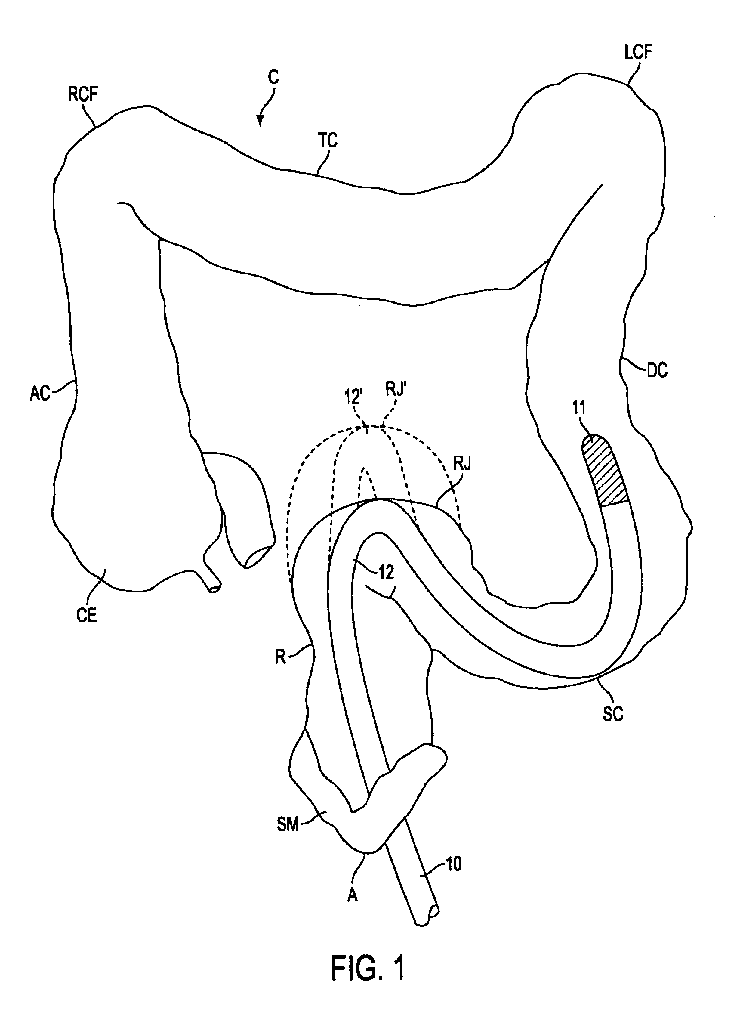
[0075]Referring to FIG. 1, problems associated with previously-known apparatus and methods for inserting and advancing a diagnostic or therapeutic instrument into a hollow body organ having tortuous or unsupported anatomy, illustratively, patient's colon C, are described. Colon C includes sphincter muscle SM disposed between anus A and rectum R. Rectum R is coupled via the rectosigmoid junction RJ to sigmoid colon SC. Sigmoid colon SC joins descending colon DC, which in turn is coupled to transverse colon TC via left colic flexure LCF. Transverse colon TC also is coupled by right colic flexure RCF to ascending colon AC and cecum CE, which receives waste products from the small intestine.

[0076]As illustrated in FIG. 1, colonoscope 10 having steerable distal tip 11 is typically inserted through anus A into rectum R, and then steered through rectosigmoid junction RJ into sigmoid colon SC. As depicted in FIG. 1, distal tip 11 of colonoscope 10 is advanced through sigmoid colon SC and de...


PUM


Abstract

Apparatus and methods are provided for placing and advancing a diagnostic or therapeutic instrument in a hollow body organ of a tortuous or unsupported anatomy, comprising a handle, an overtube disposed within a hydrophilic sheath, and a distal region having an atraumatic tip. The overtube may be removable from the handle, and have a longitudinal axis disposed at an angle relative to the handle. The sheath may be disposable to permit reuse of the overtube. Fail-safe tensioning mechanisms may be provided to selectively stiffen the overtube to reduce distension of the organ caused by advancement of the diagnostic or therapeutic instrument. The fail-safe tensioning mechanisms reduce the risk of reconfiguration of the overtube in the event that the tension system fails, and, in one embodiment, rigidizes the overtube without substantial proximal movement of the distal region. The distal region permits passive steering of the overtube caused by deflection of the diagnostic or therapeutic instrument, while the atraumatic tip prevents the wall of the organ from becoming caught or pinched during manipulation of the diagnostic or therapeutic instrument.

Description

REFERENCE TO RELATED APPLICATIONS[0001]This application is a continuation-in-part of U.S. patent application Ser. Nos. 10 / 173,203, 10 / 173,227 now U.S. Pat. No. 6,790,173, Ser. No. 10 / 173,238 now U.S. Pat. No. 6,837,847 and Ser. No. 10 / 173,220 now U.S. Pat. No. 6,783,491, all of which were filed Jun. 13, 2002 and are incorporated herein by reference in their entireties.FIELD OF THE INVENTION[0002]The present invention relates to apparatus and methods for placing and advancing a diagnostic or therapeutic instrument in a hollow body organ of unsupported anatomy, while reducing patient discomfort and risk of injury.BACKGROUND OF THE INVENTION[0003]The use of the colonoscope for examining the interior of the large intestine or colon is well-known. In general, a physician performing an examination or treatment of the colon inserts a colonoscope into the anus and then advances the colonoscope into the colon. A complete examination requires the physician to advance the colonoscope into the ...

Claims


Application Information

Patent Timeline
Patent Type & Authority Patents(United States)
IPC IPC(8): A61B1/005A61B1/01A61B1/31A61B1/32A61B1/008
CPCA61B1/0008A61B1/00082A61B1/00105A61B1/00135A61B1/00154A61B1/0052A61B1/0055A61B1/31A61B1/00078A61B1/0058A61B1/008A61B1/32
Inventor EWERS, RICHARD C.SAADAT, VAHIDREYDEL, BORIS
Owner USGI MEDICAL
Why Eureka
Try Eureka