
Multiplex analysis using membrane-bound sensitizers

a sensitizer and multi-layer technology, applied in the field of multi-layer analysis using membrane-bound sensitizers, can solve the problems of no comparable technology available for making global or system-wide measurements, and the challenge of studying such processes, and achieve the effects of convenient multiplexing capability, reduced background, and increased sensitivity

Inactive Publication Date: 2005-09-27
ACLARA BIOSCIENCES INC +1
View PDF61 Cites 66 Cited by
  • Summary
  • Abstract
  • Description
  • Claims
  • Application Information

AI Technical Summary

Benefits of technology

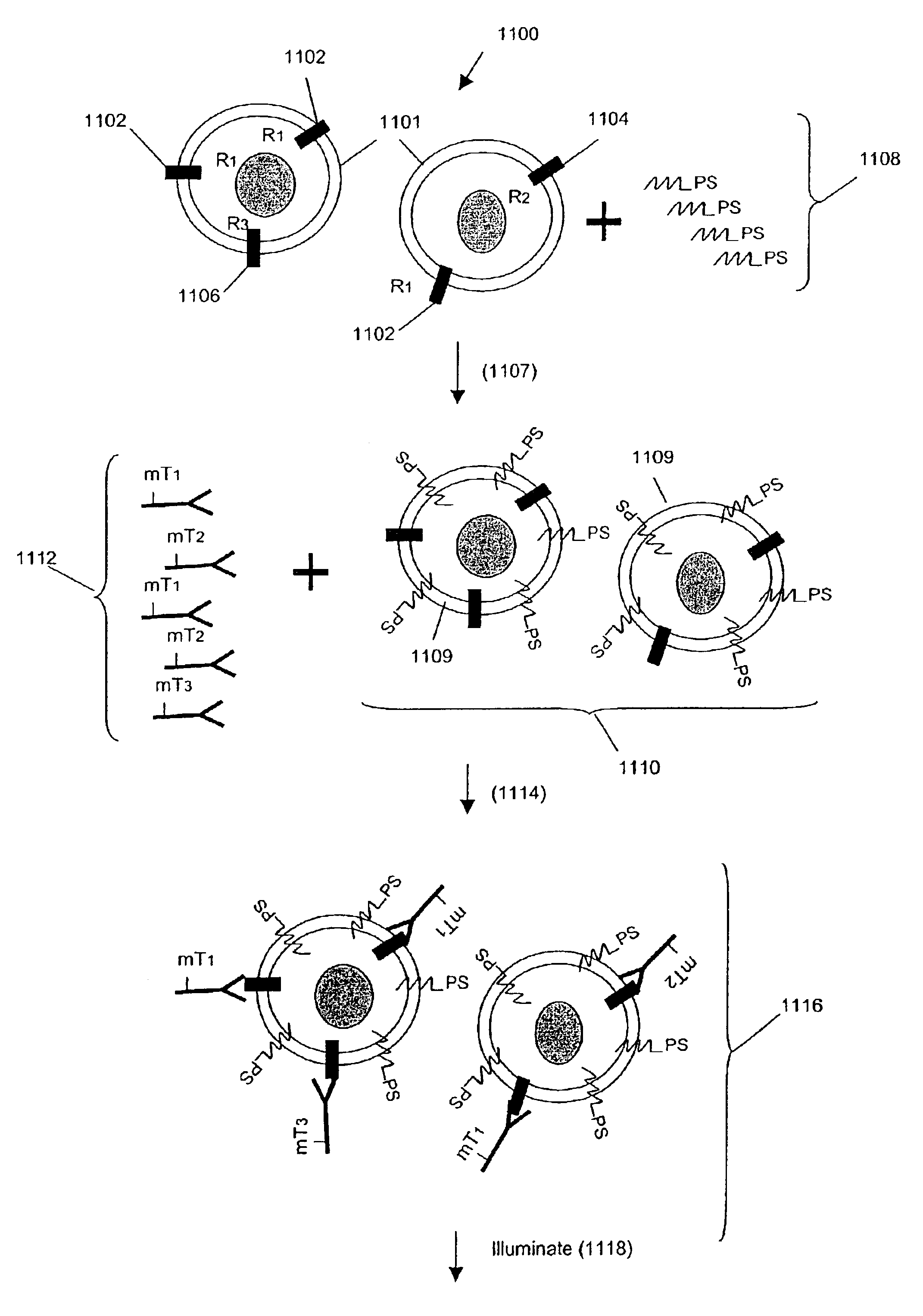
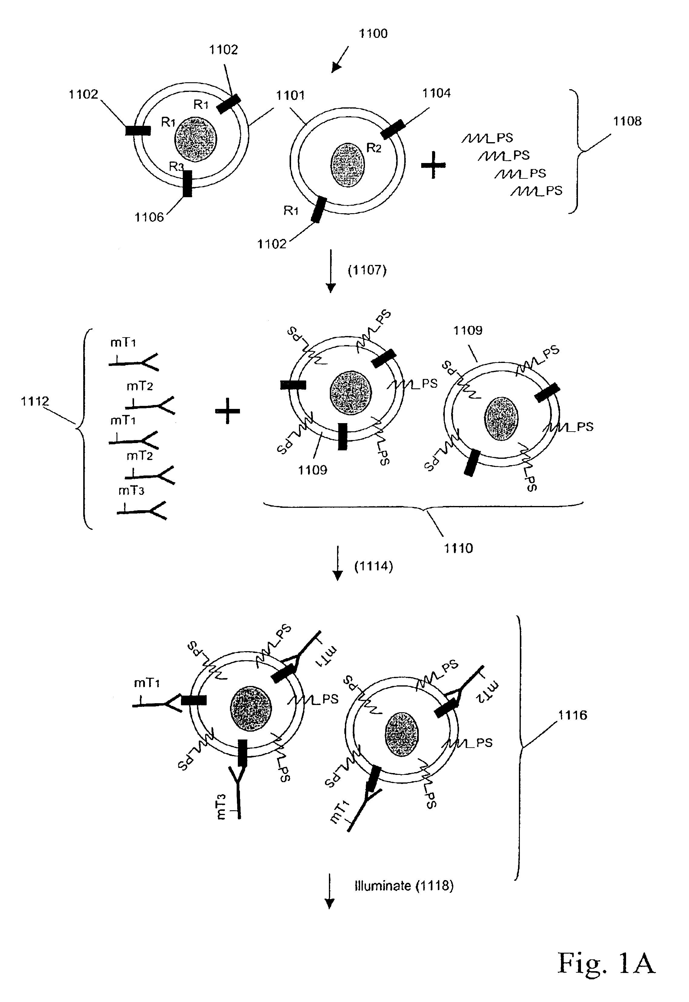
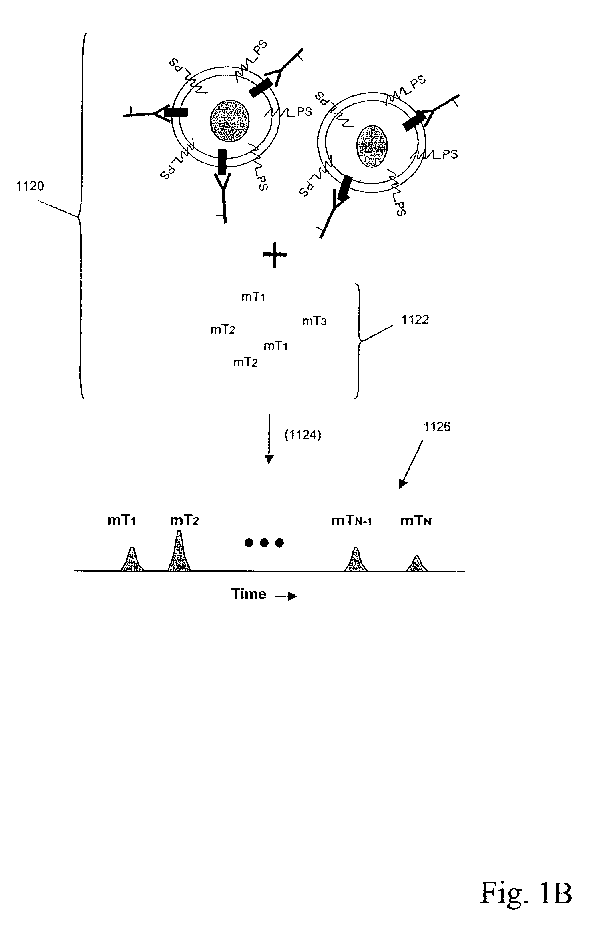
"The present invention is about a method and composition for detecting substances associated with membranes in a sample. The method involves using molecular tags that are attached to binding compounds which specifically bind to the membrane-associated substances. These molecular tags are then released upon activation of cleavage moieties, or sensitizers, anchored in the membrane. The released molecular tags are then identified using their distinct separation and detection characteristics. This method has several advantages over current techniques, including increased sensitivity and convenient multiplexing capability."

Problems solved by technology

However, studying such processes has been challenging since they are often characterized by a highly complex interaction of many molecular components, e.g. Gutkind, Science STKE: 1-13 (2000); Weng et al, Science, 284: 92-96 (1999).
However, for other phenomena, such as signal transduction pathways and other membrane-mediated processes, that involve the interaction of several to many tens of proteins, no comparable technology is available for making global or system-wide measurements.

Method used


Image

  • Multiplex analysis using membrane-bound sensitizers
  • Multiplex analysis using membrane-bound sensitizers
  • Multiplex analysis using membrane-bound sensitizers

Examples


example 1

Conjugation and Release of a Molecular Tag

[0230]FIG. 7A-B summarize the methodology for conjugation of molecular tag precursor to an antibody or other binding compound with a free amino group, and the reaction of the resulting conjugate with singlet oxygen to produce a sulfinic acid moiety as the released molecular tag. FIG. 8A-J shows several molecular tag reagents, most of which utilize 5- or 6-carboxyfluorescein (FAM) as starting material.

example 2

Preparation of Pro2, Pro4, and Pro6 through Pro13

[0231]The scheme outlined in FIG. 9A shows a five-step procedure for the preparation of the carboxyfluorescein-derived molecular tag precursors, namely, Pro2, Pro4, Pro6, Pro7, Pro8, Pro9, Pro10, Pro11, Pro12, and Pro13. The first step involves the reaction of a 5- or 6-FAM with N-hydroxysuccinimide (NHS) and 1,3-dicylcohexylcarbodiimide (DCC) in DMF to give the corresponding ester, which was then treated with a variety of diamines to yield the desired amide, compound 1. Treatment of compound 1 with N-succinimidyl iodoacetate provided the expected iodoacetamide derivative, which was not isolated but was further reacted with 3-mercaptopropionic acid in the presence of triethylamine. Finally, the resulting β-thioacid (compound 2) was converted, as described above, to its NHS ester. The various e-tag moieties were synthesized starting with 5- or 6-FAM, and one of various diamines. The diamine is given H2N^X^NH2 in the first reaction of F...

example 3

Characterization of Cell Surface Receptor Binding Using a Protein-Molecular Tag Conjugate

[0247]To demonstrate that the amount of probe tag released is related to total number of receptors in the cell sample, a ligand-receptor assay like that described in Section II above was carried out by adding a fixed amount (about 55 nM) protein probe (TNF-tag) added to samples containing increasing numbers of target cells (U937 cells) that respond to TNF. In each sample, the probe was incubated with the cells for 60 minutes, then the sample irradiated at 640-700 nm to release bound etags. The sample was separated by capillary electrophoresis, and the amount of released probe from each sample quantitated as area under the curve. In this, and the assays described below, the cells were initially modified to contain a cell-surface sensitizer agent.

[0248]The results are plotted in FIG. 17, which shows the detected level of a molecular tag, expressed as RFU (relative fluorescence units), plotted as a...


PUM

PropertyMeasurementUnit
molecular weightaaaaaaaaaa
diameteraaaaaaaaaa

Abstract

The present invention is directed to methods and compositions for determining the presence, absence, and / or amounts of one or more membrane-associated analytes in a sample. In accordance with the invention, binding compounds derivatized with releasable molecular tags specifically bind to selected membrane-associated analytes, after which the molecular tags are released upon activation of cleavage moieties, or sensitizers, anchored in the same membrane as the membrane-associated analytes. The released molecular tags are then identified by their distinct separation and detection characteristics.

Description

CROSS-REFERENCE TO RELATED APPLICATIONS AND PATENTS[0001]This application claims priority from U.S. provisional applications Ser. No. 60 / 361,975 filed Mar. 5, 2002 and Ser. No. 60 / 440,838 filed Jan. 17, 2003, which applications are incorporated herein by reference in their entirety.FIELD OF THE INVENTION[0002]This invention relates to methods and compositions for detecting and / or measuring one or more analytes embedded in or associated with lipid membranes.BACKGROUND OF THE INVENTION[0003]Many crucial biological processes take place in or adjacent to cellular membranes including receptor oligomerization, endocytosis, ligand-receptor binding, phosphorylation and de-phosphorylation of membrane proteins, folding and transport of newly synthesized proteins, viral invasion, immune reactions, and so on. Malfunctions in such processes can have profound effects on an organism's health and disease, e.g. Bunn et al, Semin. Oncol., 29 (5 Suppl 14): 38-44 (2002); Baker, Oncogene, 17: 3261-3270 ...

Claims


Application Information

Patent Timeline
Patent Type & Authority Patents(United States)
IPC IPC(8): G01N33/543G01N33/58G01N33/566G01N33/567
CPCG01N33/54306G01N33/566G01N33/58G01N33/582G01N33/567Y10T436/10C12Q1/00G01N27/00
Inventor SINGH, SHARATCHAN-HUI, PO-YING
Owner ACLARA BIOSCIENCES INC
Learn More