X-ray-tomographic imaging apparatus, X-ray-tomographic imaging method, and program

a tomographic imaging and x-ray technology, applied in the field of x-ray tomographic imaging apparatus, an x-ray tomographic imaging method, and a program, can solve the problems of deteriorating quality of reorganized tomographic image, inability for users to observe an obtained tomographic image, and inability to display the reorganized tomographic image soon after, so as to achieve efficiency and effectiveness

Inactive Publication Date: 2005-12-06
CANON KK
View PDF15 Cites 12 Cited by
  • Summary
  • Abstract
  • Description
  • Claims
  • Application Information

AI Technical Summary

Benefits of technology

[0010]Accordingly, it is an object of the present invention to obtain the image data on a tomographic section of a subject with efficiency and effectiveness.

Problems solved by technology

In this case, it is impossible for the user to observe an obtained tomographic image soon after the imaging is finished.
Therefore, it is impossible to display the reorganized tomographic image soon after the tomographic imaging is finished.
Further, since the tomographic imaging is performed a plurality of times in sequence, image information of a previous frame (electrical charges) often affects image information of the next frame, whereby the quality of the reorganized tomographic image is deteriorated.
Therefore, very large frame-memory capacity is required.

Method used


Image

  • X-ray-tomographic imaging apparatus, X-ray-tomographic imaging method, and program
  • X-ray-tomographic imaging apparatus, X-ray-tomographic imaging method, and program
  • X-ray-tomographic imaging apparatus, X-ray-tomographic imaging method, and program

Examples


first embodiment

(First Embodiment)

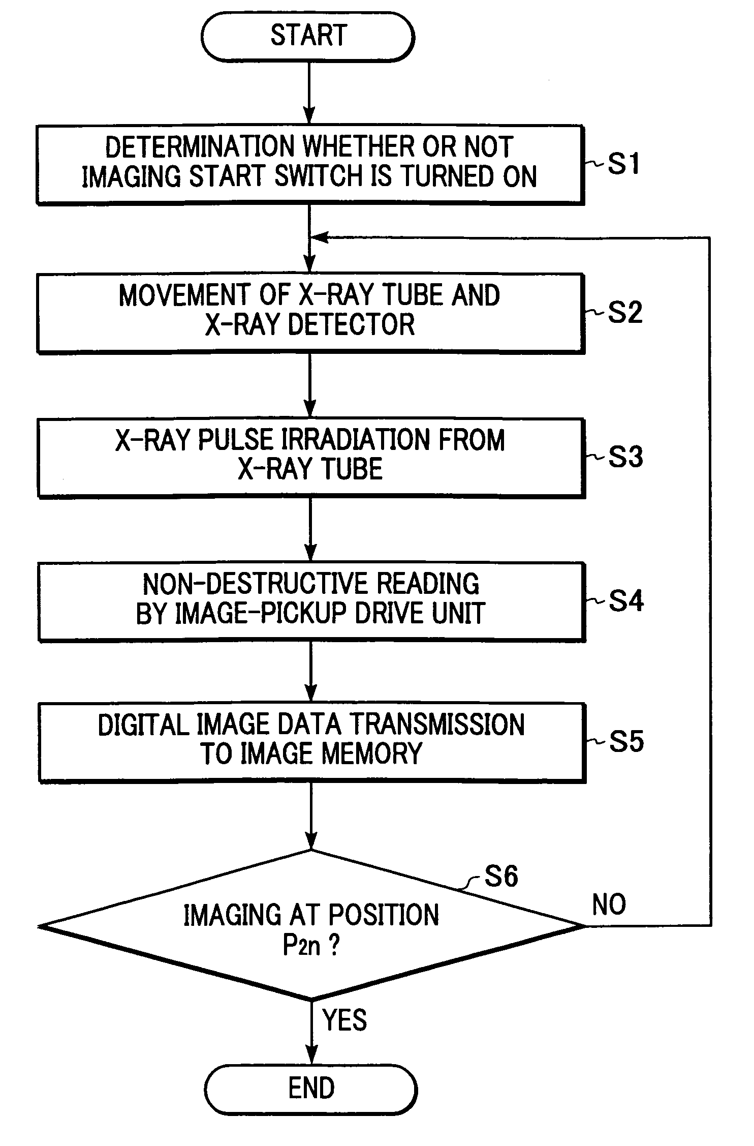
[0034]A first embodiment of the present invention will now be described with reference to FIG. 1.

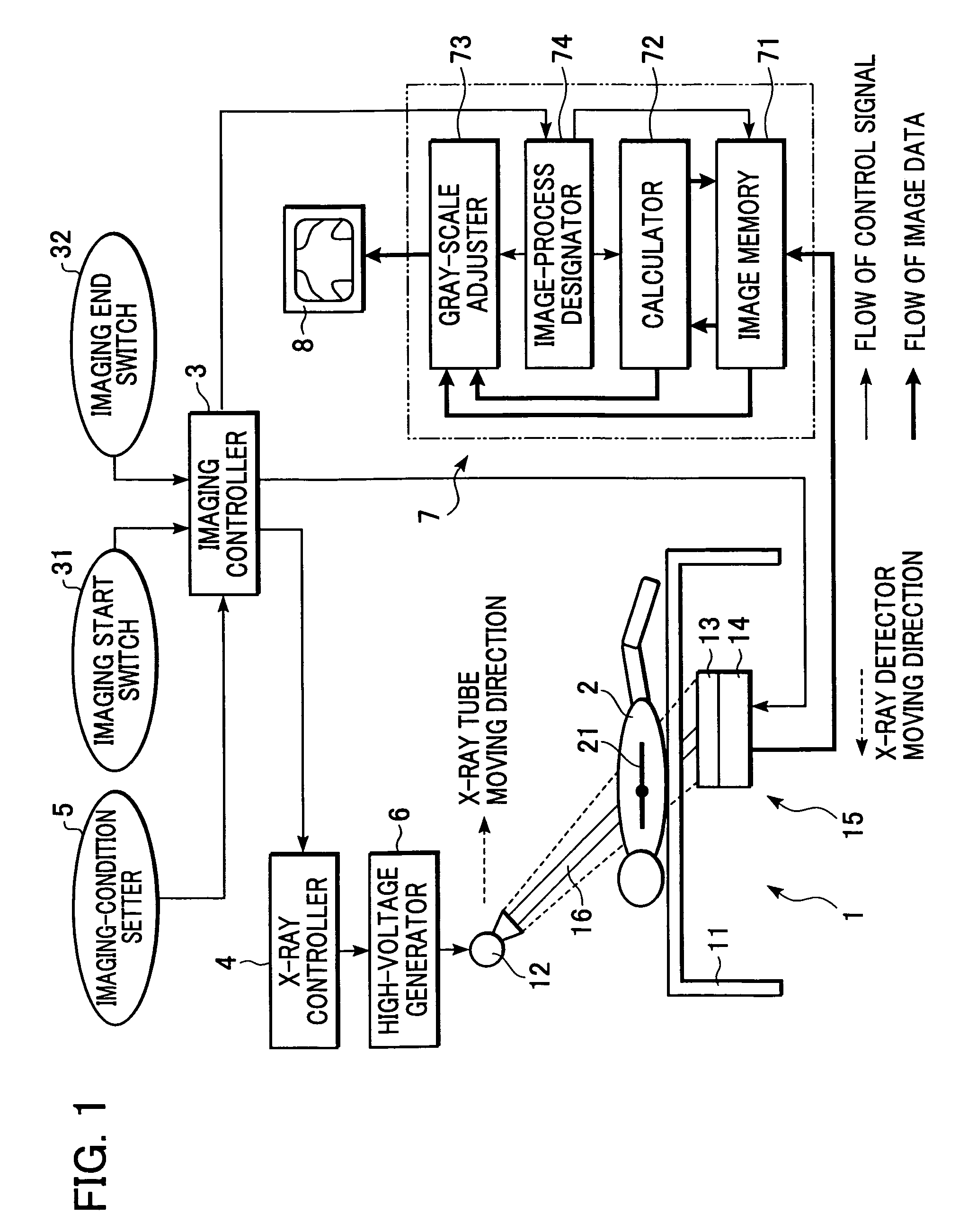
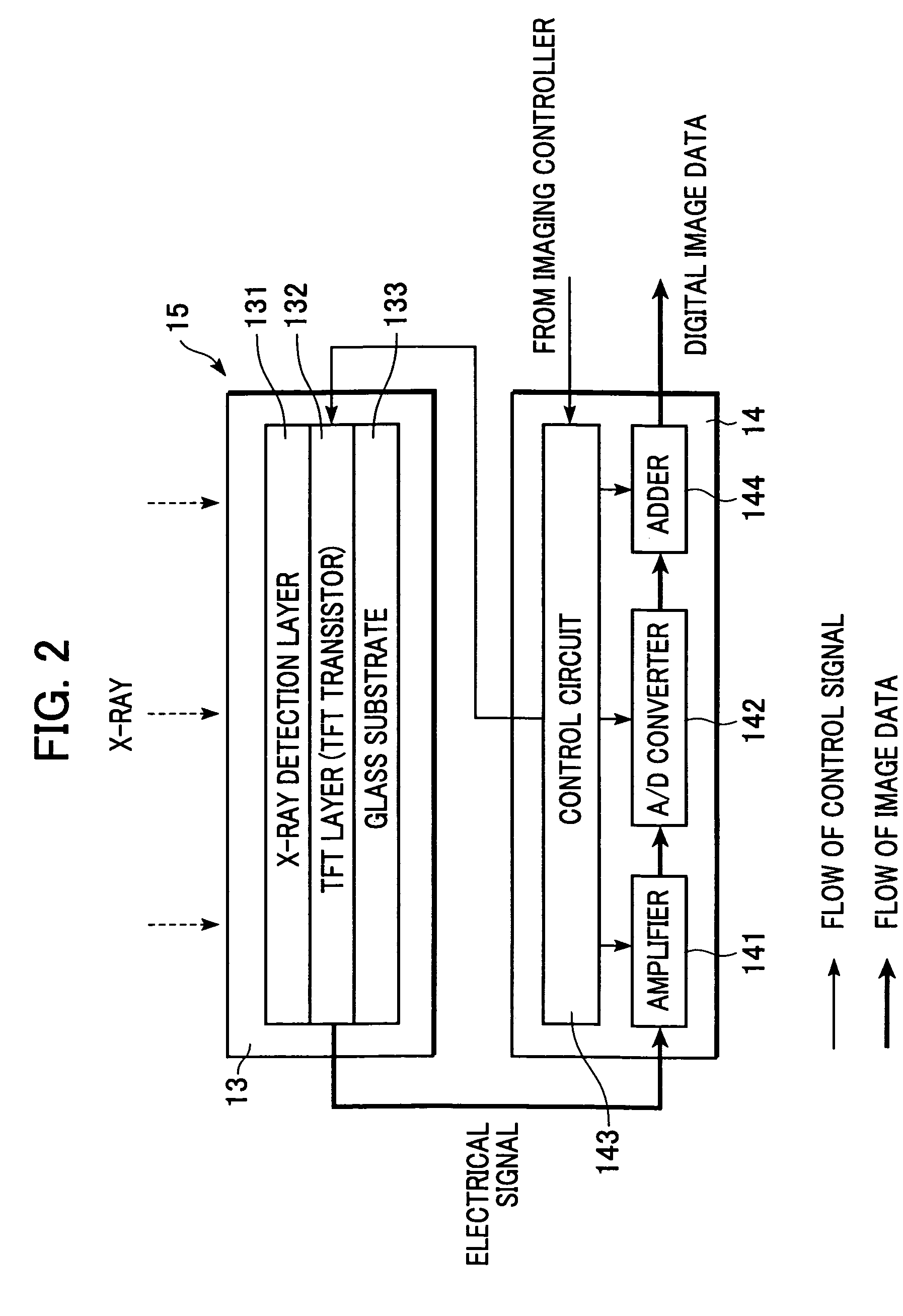
[0035]In the case of this X-ray-digital-tomographic imaging apparatus, a subject 2 is placed on a bed 11. An X-ray tube 12 is provided at a predetermined position above the bed 11. An X-ray detector 15 accommodated in a case (not shown) is provided under the bed 11. This X-ray detector 15 includes, for example, a solid-image pickup unit 13 and an image-pickup drive unit 14. This image-pickup drive unit 14 reads an X-ray image of the subject 2, as an electrical signal, by driving the solid-image pickup unit 13, and generates digital image data. The X-ray tube 12 and the X-ray detector 15 are coupled to each other by a rocking lever 16. Subsequently, the X-ray tube 12 and the X-ray detector 15 can move in parallel and in opposite directions with respect to a predetermined position on a tomographic section 21. The predetermined position is provided inside the subject 2. Th...

second embodiment

(Second Embodiment)

[0077]A second embodiment of the present invention will now be described. The configuration of an X-ray-tomographic imaging apparatus and a method of processing image data according to this embodiment is basically the same as those of the first embodiment. However, imaging conditions and a control method for performing tomographic imaging are different from those of the first embodiment.

[0078]As in the first embodiment, the operator sets the tomographic imaging conditions through the imaging-condition setter 5 as a preparation for tomographic imaging. Main conditions among these are, for example, X-ray irradiation conditions including the tube voltage, the tube current, and the X-ray irradiation time, and imaging conditions including the imaging angle range, the number of times imaging is performed, and so forth.

[0079]FIG. 8 is a schematic diagram illustrating example imaging conditions according to the second embodiment. Under these imaging conditions, the vertic...


PUM


Abstract

An X-ray-tomographic imaging apparatus for obtaining image data on a predetermined tomographic section of a subject with efficiency and effectiveness by using a plurality of X-ray projection images achieved by a plurality of X-rays that is made incident on the tomographic section from different directions is provided. The X-ray-tomographic imaging apparatus comprises a solid-image pickup unit that can convert each of the X-ray projection images to a signal and read the signal by a non-destructive reading method and a control unit making the solid-image pickup unit accumulate the signals of the X-ray projection images and read the signals by the non-destructive reading method during the signal accumulation.

Description

BACKGROUND OF THE INVENTION[0001]1. Field of the Invention[0002]The present invention relates to an X-ray-tomographic imaging apparatus, an X-ray-tomographic imaging method, and a program.[0003]2. Description of the Related Art[0004]Hitherto, X-ray-tomographic imaging has been performed by using an X-ray film. Typically, a subject is placed between an X-ray tube and the X-ray film. X-ray exposure is performed a plurality of times while the X-ray tube and the X-ray film are moved in opposite directions, with respect to the subject, in synchronization, so that the center of the X-ray beam and the center of the X-ray film are aligned. Subsequently, an X-ray-transmission image is obtained each time the X-ray exposure is performed. Further, multiple exposures of the X-ray-transmission images are performed on one film, whereby the image of a tomographic section of the subject is obtained. In this tomographic-section image, constitutional elements of the subject at a predetermined distance...

Claims


Application Information

Patent Timeline
Patent Type & Authority Patents(United States)
IPC IPC(8): A61B6/00A61B6/02G01T1/29G06T5/40H04N5/32
CPCA61B6/02A61B6/4233A61B6/461A61B6/563G01T1/2928G01T1/2985H04N5/32A61B6/025
Inventor MATSUMOTO, KAZUHIRO
Owner CANON KK
Why Eureka
Try Eureka