Device for in-vivo imaging

a technology of in-vivo imaging and imaging device, which is applied in the field of in-vivo imaging device and system, can solve the problems of low power and high bandwidth input transmitter for video signals that are unknown in the ar

Active Publication Date: 2006-03-07
GIVEN IMAGING LTD
View PDF45 Cites 647 Cited by
  • Summary
  • Abstract
  • Description
  • Claims
  • Application Information

AI Technical Summary

Benefits of technology

[0005]The device and system of the present invention enable obtaining in vivo images from within body lumens or cavities, such as images of the entire length of the gastrointestinal (GI) tract. The device and system contain an imaging system that includes a complementary metal oxide semiconductor (CMOS) imaging camera. The device also contains an ultra low power radio frequency (RF) transmitter for transmitting signals from the CMOS imaging camera to a receiving system.

Problems solved by technology

Further, an ultra low power, high bandwidth input transmitter for video signals is unknown in the art.

Method used


Image

  • Device for in-vivo imaging
  • Device for in-vivo imaging
  • Device for in-vivo imaging

Examples


Embodiment Construction

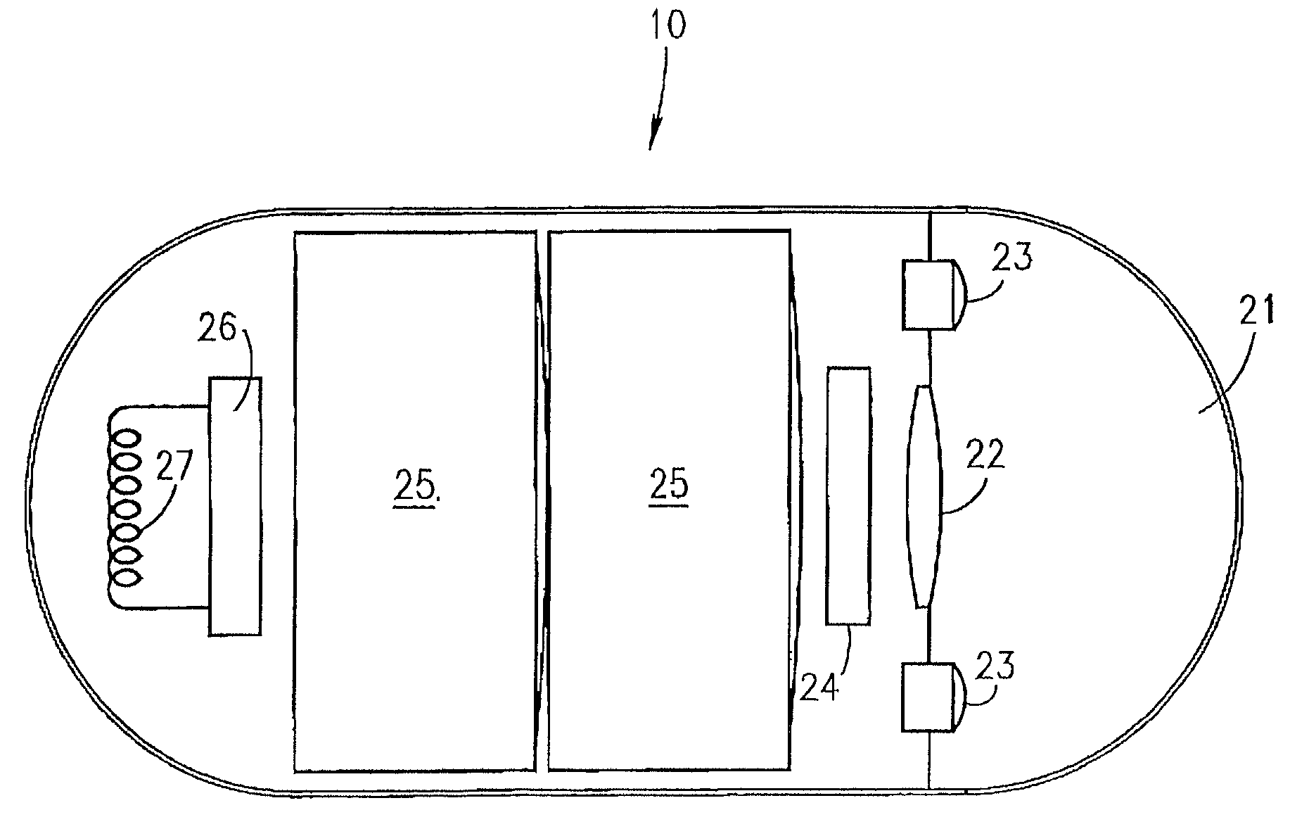
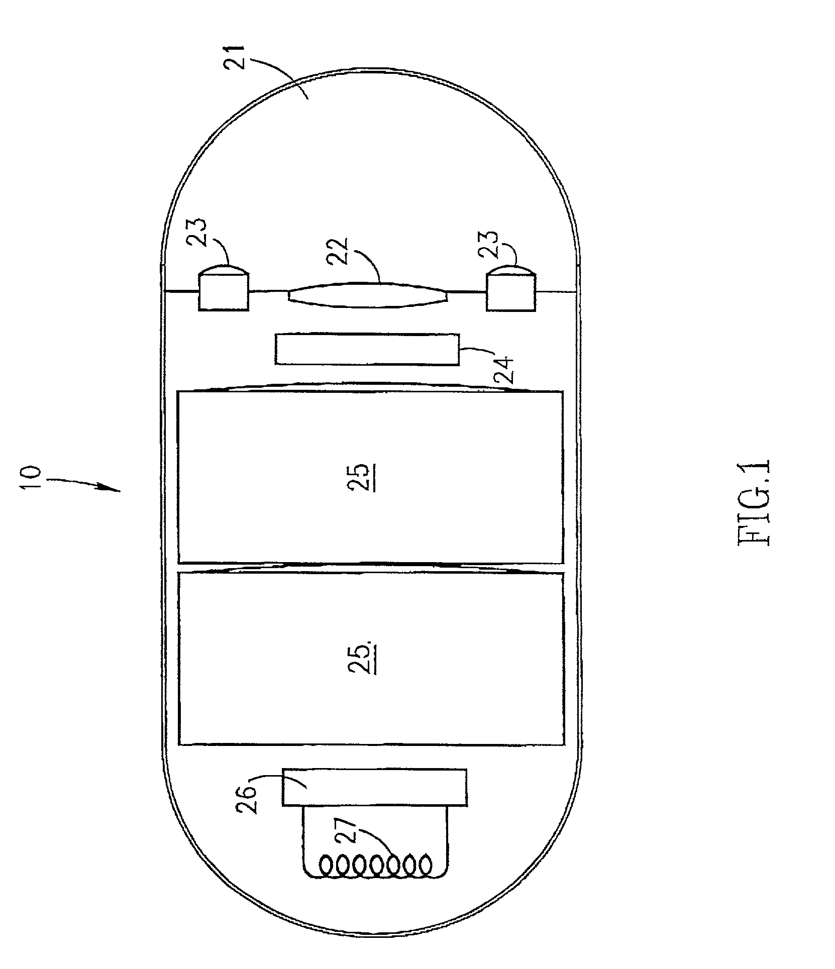
[0029]The device and system of the invention are utilized for viewing inside body lumens and cavities and for transmitting at least video data.

[0030]Reference is now made to FIG. 1 which illustrates the device and its components, according to an embodiment of the invention. The device 10 typically comprises an optical window 21 and an imaging system for obtaining images from inside a body lumen, such as the GI tract. The imaging system includes an illumination source 23, such as a white LED, a CMOS imaging camera 24, which detects the images and an optical system 22 which focuses the images onto the CMOS imaging camera 24. The illumination source 23 illuminates the inner portions of the body lumen through optical window 21. Device 10 further includes a transmitter 26 and an antenna 27 for transmitting the video signal of the CMOS imaging camera 24, and a power source 25, such as a silver oxide battery, that provides power to the electric elements of the device 10.

[0031]It will be ap...


PUM


Abstract

The present invention provides a system and method for obtaining in vivo images. The system contains an imaging system and an ultra low power radio frequency transmitter for transmitting signals from the CMOS imaging camera to a receiving system located outside a patient. The imaging system includes at least one CMOS imaging camera, at least one illumination source for illuminating an in vivo site and an optical system for imaging the in vivo site onto the CMOS imaging camera.

Description

CROSS-REFERENCE TO RELATED APPLICATIONS[0001]This application claims priority from U.S. provisional patent application No. 60 / 187,883, filed Mar. 8, 2000, entitled “In Vivo Imaging Device and System”, which is hereby incorporated by reference in its entirety.FIELD OF THE INVENTION[0002]The present invention relates to an in vivo imaging device and system such as for imaging the digestive tract.BACKGROUND OF THE INVENTION[0003]Among known in vivo measuring systems are endoscopes, which are often utilized to provide images of the upper or lower gastro-intestinal tract. However, endoscopes do not provide views of the entire length of the small intestines. Additionally, they are uncomfortable, may cause damage to the patient and are complex to operate.[0004]Swallowable electronic capsules which are moved through the digestive tract through the action of digestion and which collect data and transmit the data to a receiver system are known, one such example is the “Heidelberg” capsule. Ye...

Claims


Application Information

Patent Timeline
Patent Type & Authority Patents(United States)
IPC IPC(8): H04N7/18A61B1/04H04N9/47G03B15/00A61B1/00A61B1/05A61B1/273A61B1/31A61B5/00A61B5/07G03B15/02H04L25/03H04N5/225
CPCA61B1/00016A61B1/00036A61B1/041A61B1/0607A61B1/0676A61B1/0684A61B5/0031A61B5/073A61B5/14539H04L25/03866H04N5/2251H04N5/2252H04N5/2256H04N5/23203H04N7/185H04N2005/2255A61B1/2733A61B1/2736A61B1/31A61B2560/0209A61B1/00032H04N23/51H04N23/56H04N23/555H04N23/66H04N23/50A61B1/0016
Inventor IDDAN, GAVRIEL J.AVNI, DOVGLUKHOVSKY, ARKADYMERON, GAVRIEL
Owner GIVEN IMAGING LTD
Why Eureka
Try Eureka