Cone beam CT scanners with reduced scan length

a ct scanner and cone beam technology, applied in the direction of instruments, patient positioning for diagnostics, applications, etc., to achieve the effect of reducing the number of rotations, increasing the scan pitch, and reducing the scan length

Inactive Publication Date: 2006-03-28
PHILIPS MEDICAL SYST TECH
View PDF15 Cites 16 Cited by
  • Summary
  • Abstract
  • Description
  • Claims
  • Application Information

AI Technical Summary

Benefits of technology

[0014]In modem helical CT scanners the number of slices and hence the pitch of the scan is increased. This results in a marked reduction in the number of rotations of the X-ray tube that are required. In many cases, about one rotation is sufficient. Under these circumstances, the useless exposure of regions that are not contained in a desired volumetric region of interest (VROI) can be a significant portion of the total radiation exposure of the patient. Reduction in the scan length by even a fraction of a rotation, while still imaging the desired VROI, can result in a significant overall reduction in the patient radiation exposure.

Problems solved by technology

Under these circumstances, the useless exposure of regions that are not contained in a desired volumetric region of interest (VROI) can be a significant portion of the total radiation exposure of the patient.

Method used


Image

  • Cone beam CT scanners with reduced scan length
  • Cone beam CT scanners with reduced scan length
  • Cone beam CT scanners with reduced scan length

Examples


Embodiment Construction

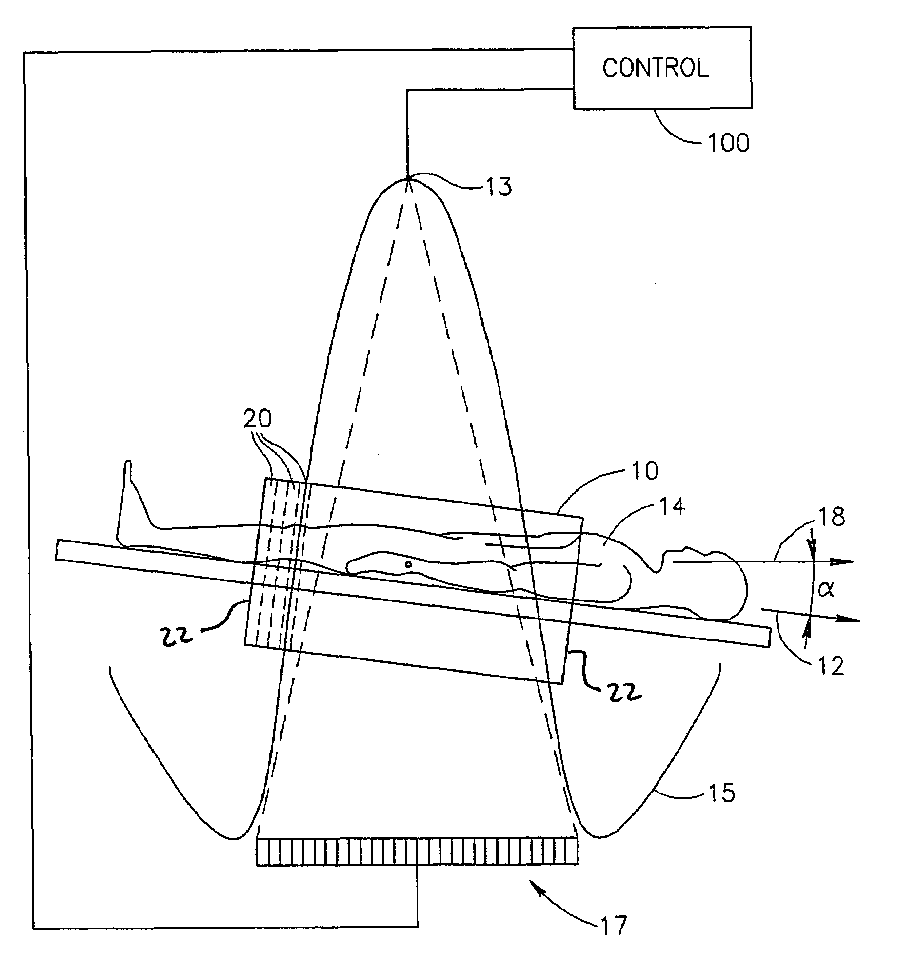
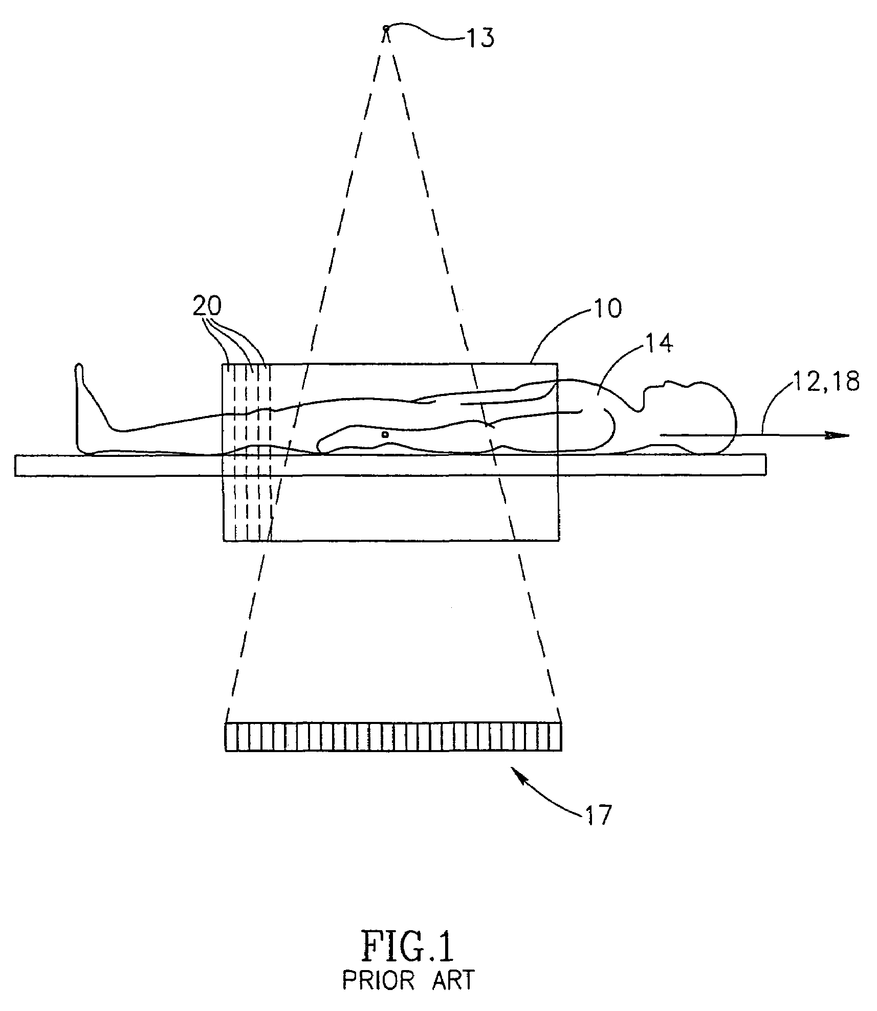
[0048]FIG. 1 shows a side view illustrating the geometry of a multi-slice cone beam according to the prior art. A cylindrical volumetric region of interest (VROI) 10 having a patient axis 12 is scanned by an X-ray source 13, while rotating about a rotation (CT) axis 18, which is shown as being coincident with axis 12 for the prior art system shown. A patient 14 moves in a direction 12. Marked on VROI 10 are a series of slices 20 (only a few of which are indicated), for which images are desired. In the prior art system, when transaxial slices (as shown) are to be imaged, the patient axis and the CT axis are coincident.

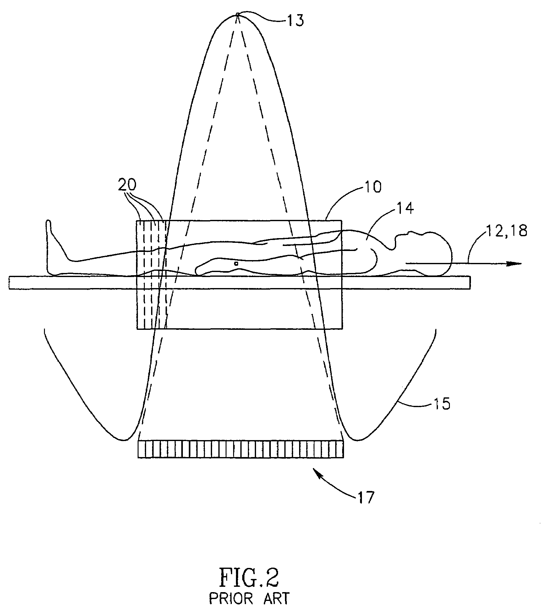
[0049]For the purposes of description of the present invention, the presentation of FIG. 2, which is equivalent to that of FIG. 1, is useful. While in FIG. 1, the patient is considered to have the motion along its axis and the X-ray source is considered to have a pure rotational motion, in FIG. 2, source 13 is shown as describing a helical motion 15 and the patient is c...


PUM


Abstract

Computerized tomography apparatus for reconstructing attenuation values within a volume comprising: an x-ray source situated operative to rotate about said volume, in a rotation plane, while irradiating at least a portion of the volume; a plurality of rows of x-ray detectors illuminated by said rotating x-ray source situated on an opposite side of the volume; a patient support operative to move a patient through a space between the source and detectors at an angle to the normal to the rotation plane, while the x-ray source illuminates the detectors; and a controller operative to compute the angle based on at least one of a radius R of said rotation, a radius r of said volume and a helix pitch, m, defined as a distance the patient support moves during a single rotation of the x-ray source.

Description

FIELD OF THE INVENTION[0001]The invention relates to CT helical scanners utilizing a cone beam source.BACKGROUND OF THE INVENTION[0002]An important criterion in the performance of computed tomography (CT) is the ability to image a large volume in a short time period. This allows for reduced artifacts caused by patient movement and for higher patient throughput.[0003]One method of improving throughput is to utilize a helical scan. In such a scan, an x-ray source that irradiates a patient rotates about the patient in a helical trajectory. Attenuation data is acquired along the entire trajectory. This data is used to reconstruct “slices” of cross-sectional attenuation images perpendicular to the axis of the helix.[0004]Another method, sometimes used in conjunction with the helix scan, provides a plurality of axially displaced rows of circumferentially spaced detectors. Scanners embodying this method are sometimes called “multi-slice” scanners. Generally, a single source is used to irra...

Claims


Application Information

Patent Timeline
Patent Type & Authority Patents(United States)
IPC IPC(8): A61B6/03A61B6/04
CPCA61B6/0457A61B6/027A61B6/4447A61B6/0487
Inventor SHECHTER, GILAD
Owner PHILIPS MEDICAL SYST TECH
Why Eureka
Try Eureka