CT and X-ray luminescence computed dual-mode synchronous tomography method

A technology of excitation fluorescence and tomography, which is applied in the fields of radiological diagnosis instruments, diagnosis, medical science, etc., and can solve the problems of high dose, long time, and complicated process.

Active Publication Date: 2016-07-27
FOURTH MILITARY MEDICAL UNIVERSITY
View PDF3 Cites 8 Cited by
  • Summary
  • Abstract
  • Description
  • Claims
  • Application Information

AI Technical Summary

Problems solved by technology

[0005] The purpose of the present invention is to provide a dual-mode simultaneous tomographic imaging method of CT and X-ray excited fluorescence, aiming to solve the problems of separate acquisition, complicated process, high dose and long time of CT / XLCT dual-mode imaging

Method used


Image

  • CT and X-ray luminescence computed dual-mode synchronous tomography method
  • CT and X-ray luminescence computed dual-mode synchronous tomography method
  • CT and X-ray luminescence computed dual-mode synchronous tomography method

Examples


Embodiment Construction

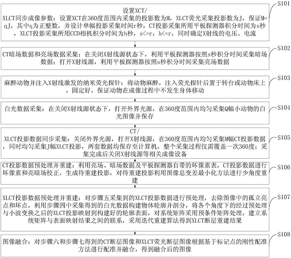
[0042] In order to make the object, technical solution and advantages of the present invention more clear, the present invention will be further described in detail below in conjunction with the examples. It should be understood that the specific embodiments described here are only used to explain the present invention, not to limit the present invention.

[0043] The application principle of the present invention will be further described below in conjunction with specific embodiments.

[0044] The CT / XLCT dual-mode imaging system of our laboratory was used to simultaneously acquire the CT and XLCT projection data of the phantom. The X-ray source of the system is the micro-focus spot X-ray source of Oxford Instruments (UltraBright, Oxford Instruments, and U.K.), the X-ray flat panel detector is Dexela2923 (Dexela2923CMOSX-raydetector), and the CCD camera adopts electron multiplying CCD (iXonDU-897 , Andor, United Kingdom). The system uses the rotation of the turntable to dr...


PUM


Abstract

The invention discloses a CT and X-ray luminescence computed dual-mode synchronous tomography method. Small animal X-ray luminescence computed tomography and computed tomography (CT) can be achieved synchronously, and a more accurate imaging object anatomical structure and molecular imaging information are provided. It is guaranteed that an XLCT projection and a CT projection are collected at the same location and same time by reducing the CT projection angle number and setting the CT projection angle number to be the same as that of the XLCT or be in a diploid relation with that of the XLCT; meanwhile, by means of a compressive sensing principle, a few-angle reconstruction algorithm achieving image total variation minimization is adopted for the CT projection, and a precondition matrix and wavelet transformation are introduced into XLCT iteration reconstruction so as to reduce ill conditioning of reconstruction. According to the CT and X-ray luminescence computed dual-mode synchronous tomography method, synchronous tomography of CT / XLCT can be achieved, dual-mode tomography processes are simplified, the experimental difficulty is reduced, the imaging time is reduced, the experimental failure rate is decreased, a radiation dosage is decreased, an error caused by objecting moving in the imaging process is avoided, and image registration and fusion difficulties are reduced.

Description

technical field [0001] The invention belongs to the interdisciplinary fields of physics mathematics, biomedical engineering and computer, and in particular relates to a dual-mode synchronous tomographic imaging method of CT and X-ray excited fluorescence. Background technique [0002] X-ray computed tomography (Computed Tomography, CT) has been widely used in high-resolution anatomical structure imaging of small animals, but its application fields are limited due to the lack of biomolecular imaging information. In recent years, with the development of X-ray nanofluorescent probes, X-ray Luminescence Computed Tomography (XLCT), as a new method of molecular tomography that combines X-ray and optical imaging, is expected to realize deep tumor in vivo Position marking, tracking and judgment of tumor size. CT / XLCT dual-mode imaging, both of which rely on X-rays, the former can provide information on the anatomical structure of small animals, and the latter can provide image info...

Claims


Application Information

Patent Timeline
Patent Type & Authority Applications(China)
IPC IPC(8): A61B6/00
CPCA61B6/00A61B6/4417A61B6/482A61B6/5241
Inventor 戎军艳卢虹冰高鹏廖琪梅刘文磊刘洋见伟平
Owner FOURTH MILITARY MEDICAL UNIVERSITY
Why Eureka
Try Eureka