Method and apparatus for three-dimensional filtering of angiographic volume data

a volume data and filtering technology, applied in the field of three-dimensional angiography, can solve the problems of complex analysis and interpretation of unprocessed gray scale angiographic images, degrade quality of life, and increase complexity, so as to achieve rapid filtering and remove confusing and obscuring non-vascular contrast.

Inactive Publication Date: 2006-04-04
KONINKLIJKE PHILIPS ELECTRONICS NV +1
View PDF25 Cites 77 Cited by
  • Summary
  • Abstract
  • Description
  • Claims
  • Application Information

AI Technical Summary

Benefits of technology

[0016]One advantage of the present invention is that it substantially removes confusing and obscuring non-vascular contrast from an angiographic image.
[0017]Another advantage of the present invention is that it accounts during the filtering for the range of vessel size scales which are typically present in angiographic data.
[0018]Yet another advantage of the

Problems solved by technology

Catastrophic medical events such as heart attacks and strokes that result from underlying vascular problems are a leading cause of death in the United States.
Many Americans also suffer from chronic vascular diseases which degrade quality of life.
Analysis and interpretation of the unprocessed gray scale angiographic image is complicated by a number of factors.
Complexity arises because blood vessel networks in the human body are highly intricate, and a particular image will typically include tortuous or occluded blood vessels, shape variability, regions of very high blood vessel densities, a wide range of blood vessel diameters, and the like.
Additionally, angiographic techniques, although designed to selectively image the va

Method used


Image

  • Method and apparatus for three-dimensional filtering of angiographic volume data
  • Method and apparatus for three-dimensional filtering of angiographic volume data
  • Method and apparatus for three-dimensional filtering of angiographic volume data

Examples


Embodiment Construction

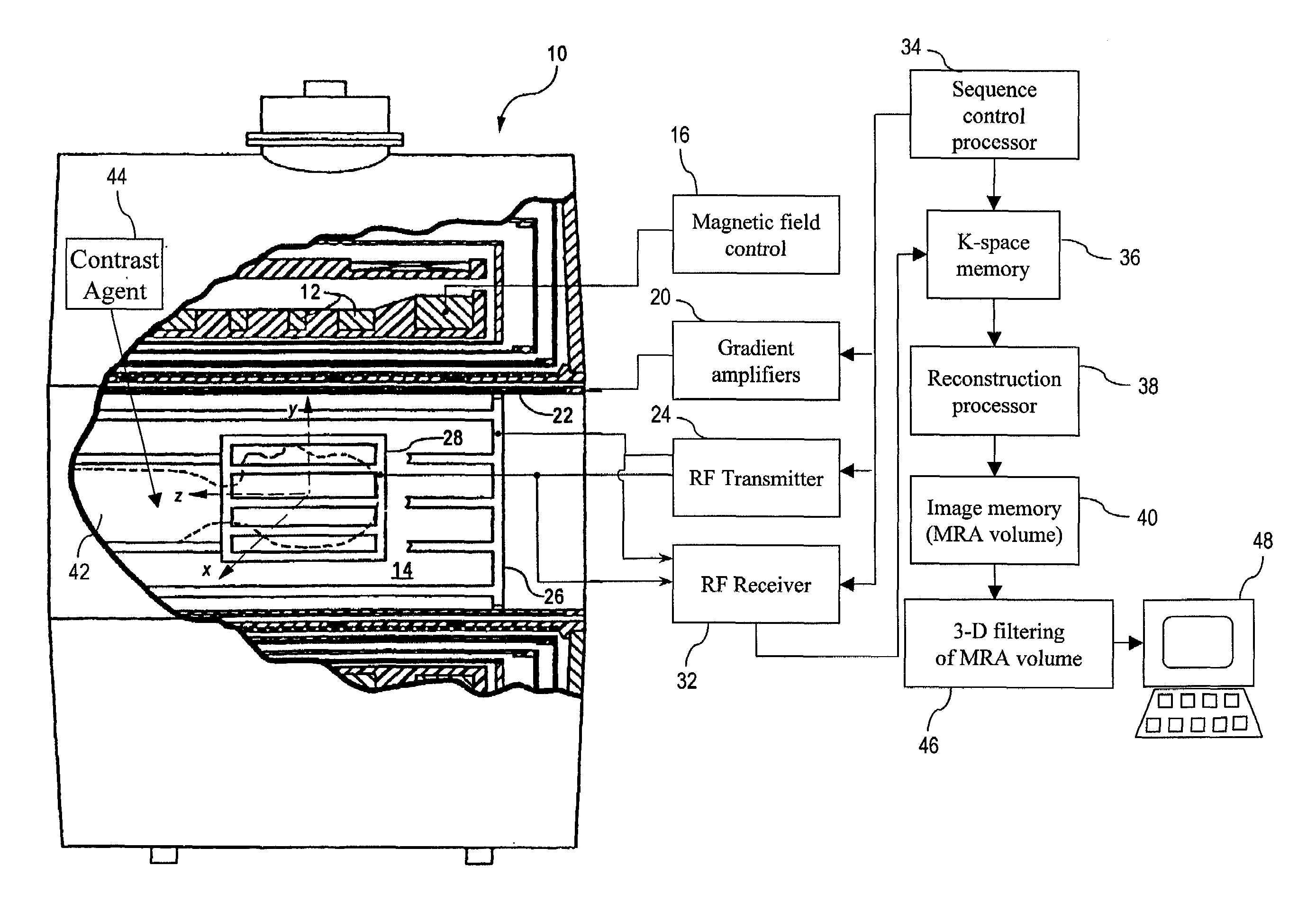
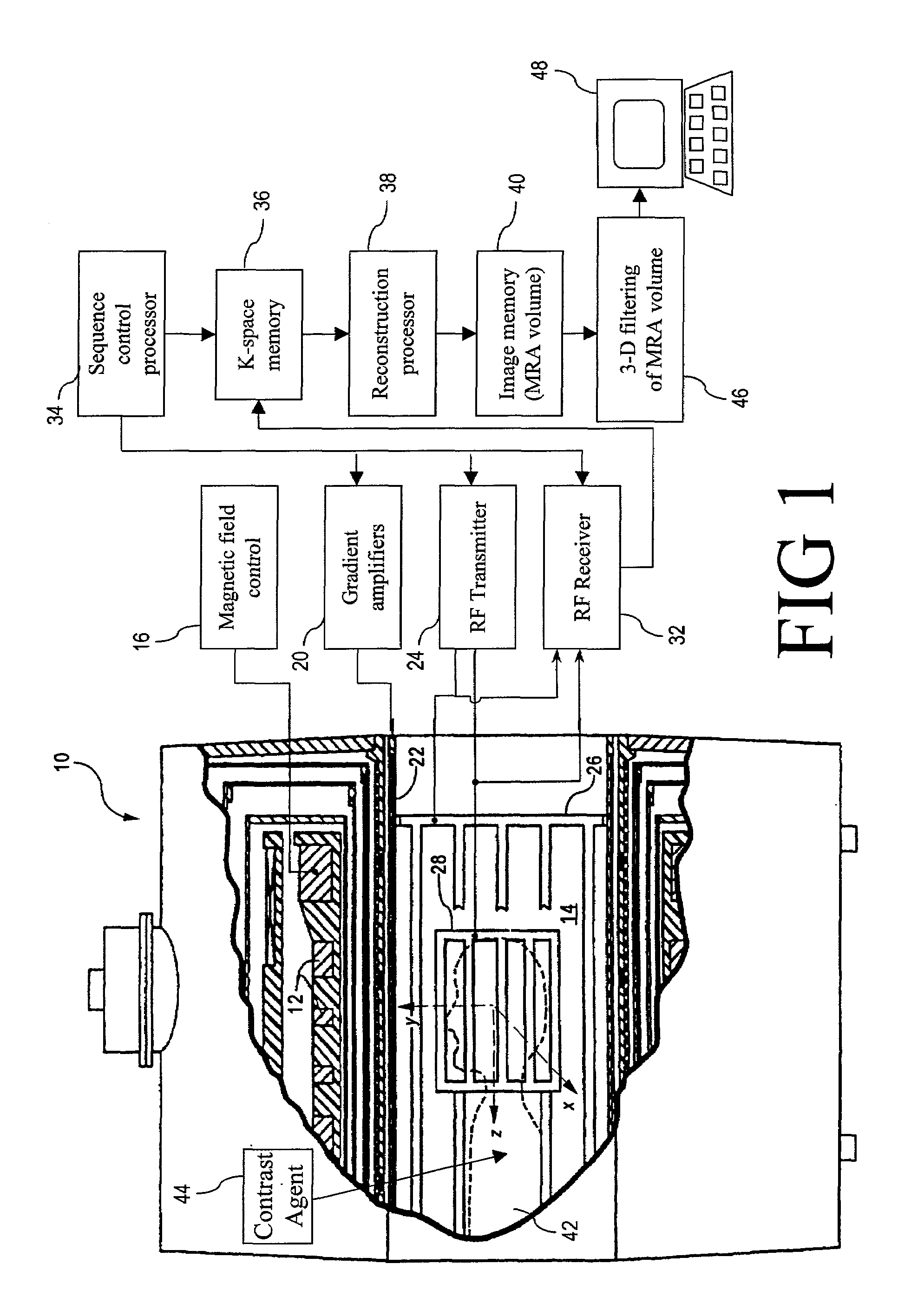
[0040]With reference to FIG. 1, a magnetic resonance imaging system that suitably practices volumetric angiographic imaging in accordance with an embodiment of the invention is described. Although the invention is described herein with respect to a magnetic resonance imaging embodiment, those skilled in the art will appreciate that the invention is applicable to a broad range of three-dimensional angiographic modalities and techniques, including but not limited to contrast-enhanced magnetic resonance angiography, non-contrast enhanced magnetic angiography, and computed tomographic angiography.

[0041]With reference to FIG. 1, a magnetic resonance imaging (MRI) scanner 10 typically includes superconducting or resistive magnets 12 that create a substantially uniform, temporally constant main magnetic field B0 along a z-axis through an examination region 14. Although a bore-type magnet is illustrated in FIG. 1, the present invention is equally applicable to open magnet systems and other ...


PUM


Abstract

A filtering method and apparatus for selectively filtering a gray scale angiographic image volume (54) that images vascular structures and that also contains extraneous non-vascular contrast is disclosed. A minimum vessel width parameter Wmin (70) is selected. A maximum vessel width parameter Wmax (132) is selected. A range of widths (66) are selected such that each vessel width parameter Wi (194) lies between Wmin (70) and Wmax (132) inclusive. For each Wi (194), an image Ii(x,y,z) (198) is generated that selectively retains imaged vascular structures of diameter Wi (194). The images Ii(x,y,z) (198) are combined to form a filtered image IF(x,y,z) (64) that selectively images the vascular structures.

Description

BACKGROUND OF THE INVENTION[0001]The present invention relates to the medical imaging arts. It particularly relates to three-dimensional angiography using the magnetic resonance imaging (MRI) and computed tomography (CT) medical imaging techniques, and will be described with particular reference thereto. However, the invention will also find application in conjunction with other three-dimensional or two-dimensional angiographic imaging modalities as well as in other imaging arts in which tubular structures and networks of non-uniform diameter are advantageously differentiated from extraneous imaged structures and background noise.[0002]Catastrophic medical events such as heart attacks and strokes that result from underlying vascular problems are a leading cause of death in the United States. Many Americans also suffer from chronic vascular diseases which degrade quality of life.[0003]Angiography, which relates to the imaging of blood vessels and blood vessel systems, is a powerful m...

Claims


Application Information

Patent Timeline
IPC IPC(8): G06K9/00G01R33/56G01R33/563G06T17/00
CPCG06T17/00G01R33/563G01R33/5608
Inventor SURI, JASJIT S.LIU, KECHENG
Owner KONINKLIJKE PHILIPS ELECTRONICS NV
Why Eureka
Try Eureka