

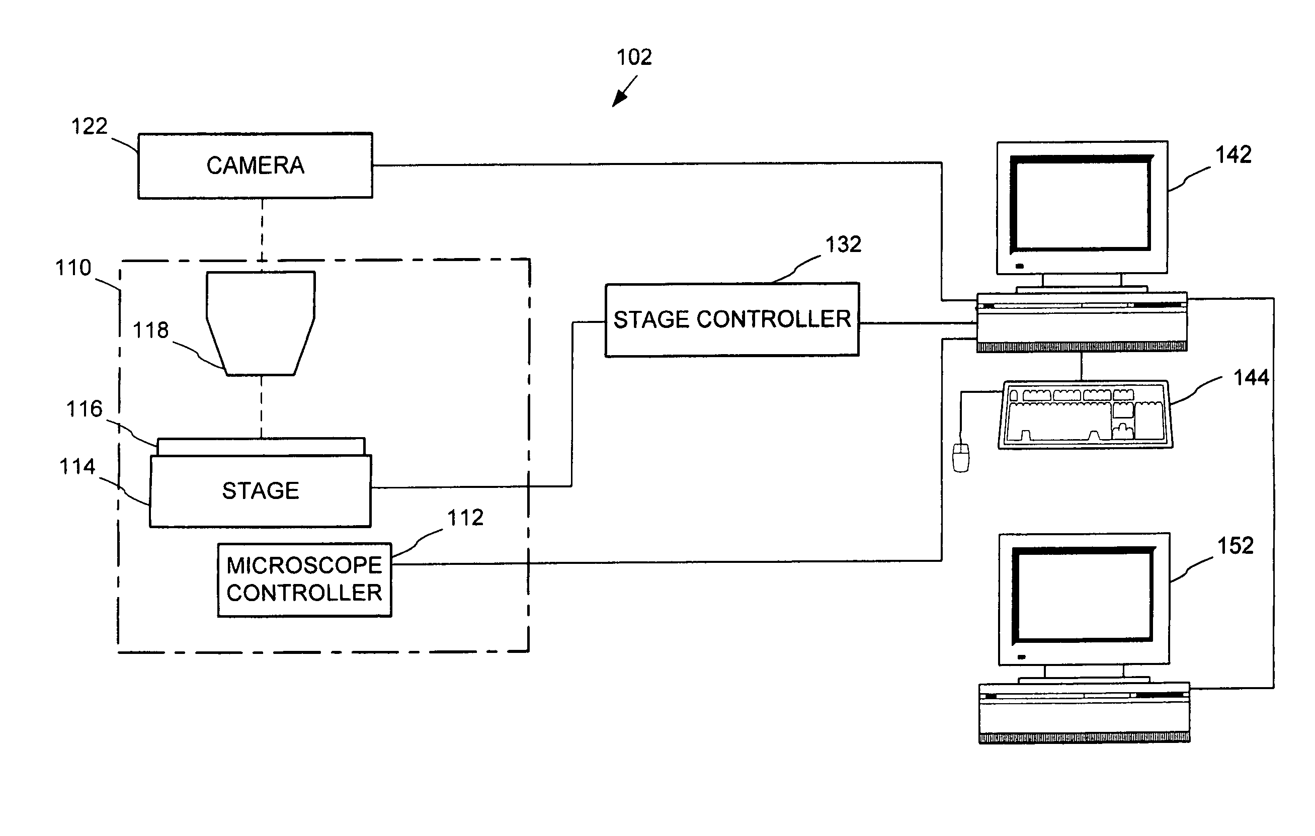
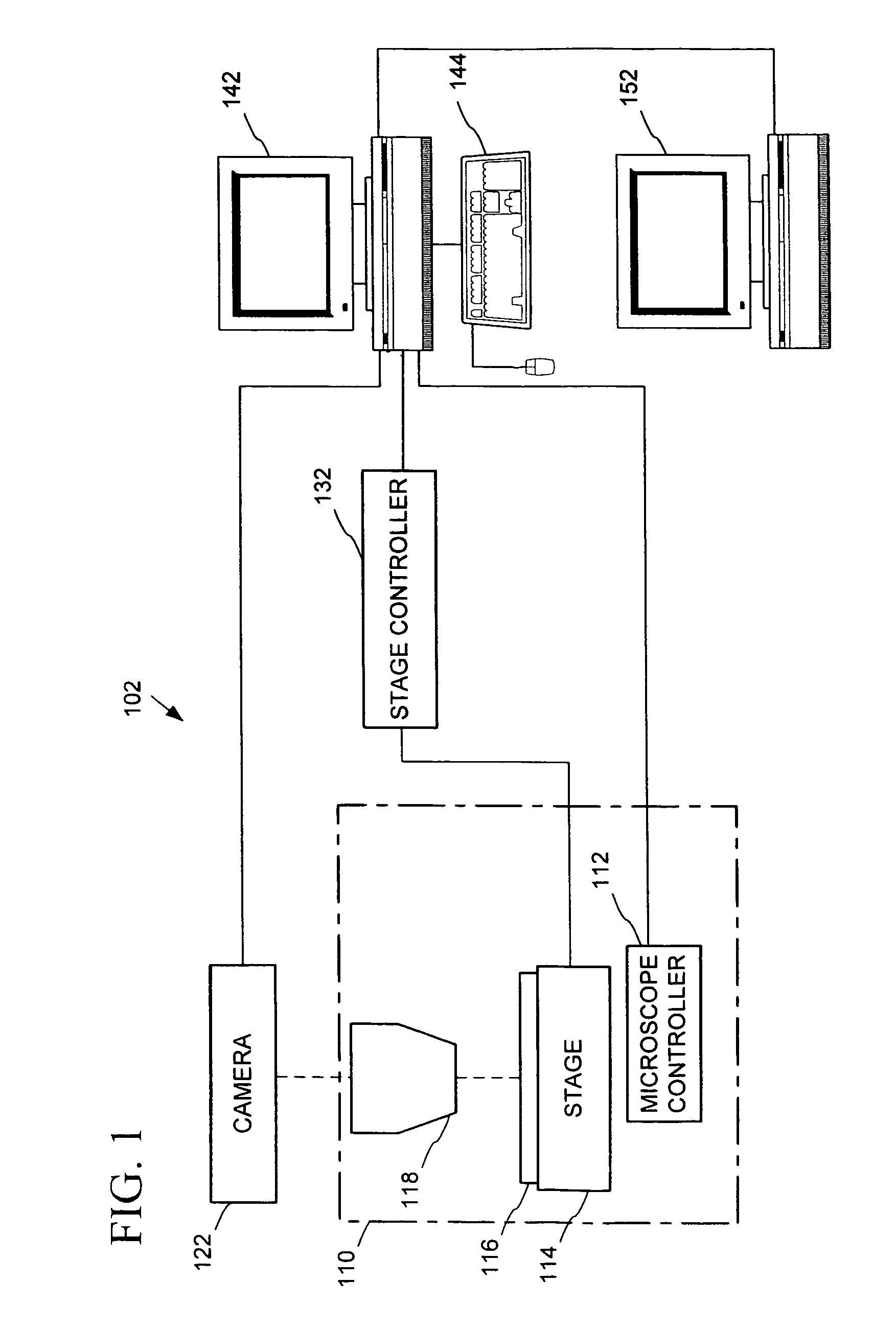
Automated microscopic image acquisition, compositing, and display
a technology of automatic acquisition and microscopic image, applied in image enhancement, image analysis, instruments, etc., can solve the problems of difficult to find such exemplary biological samples, difficulty in acquiring such a set of training slides, and deterioration, so as to avoid errors due to variations in focal plane, avoid delay in browsing images, and avoid delay
- Summary
- Abstract
- Description
- Claims
- Application Information
AI Technical Summary
Benefits of technology
Problems solved by technology
Method used
Image

Examples
example implementation
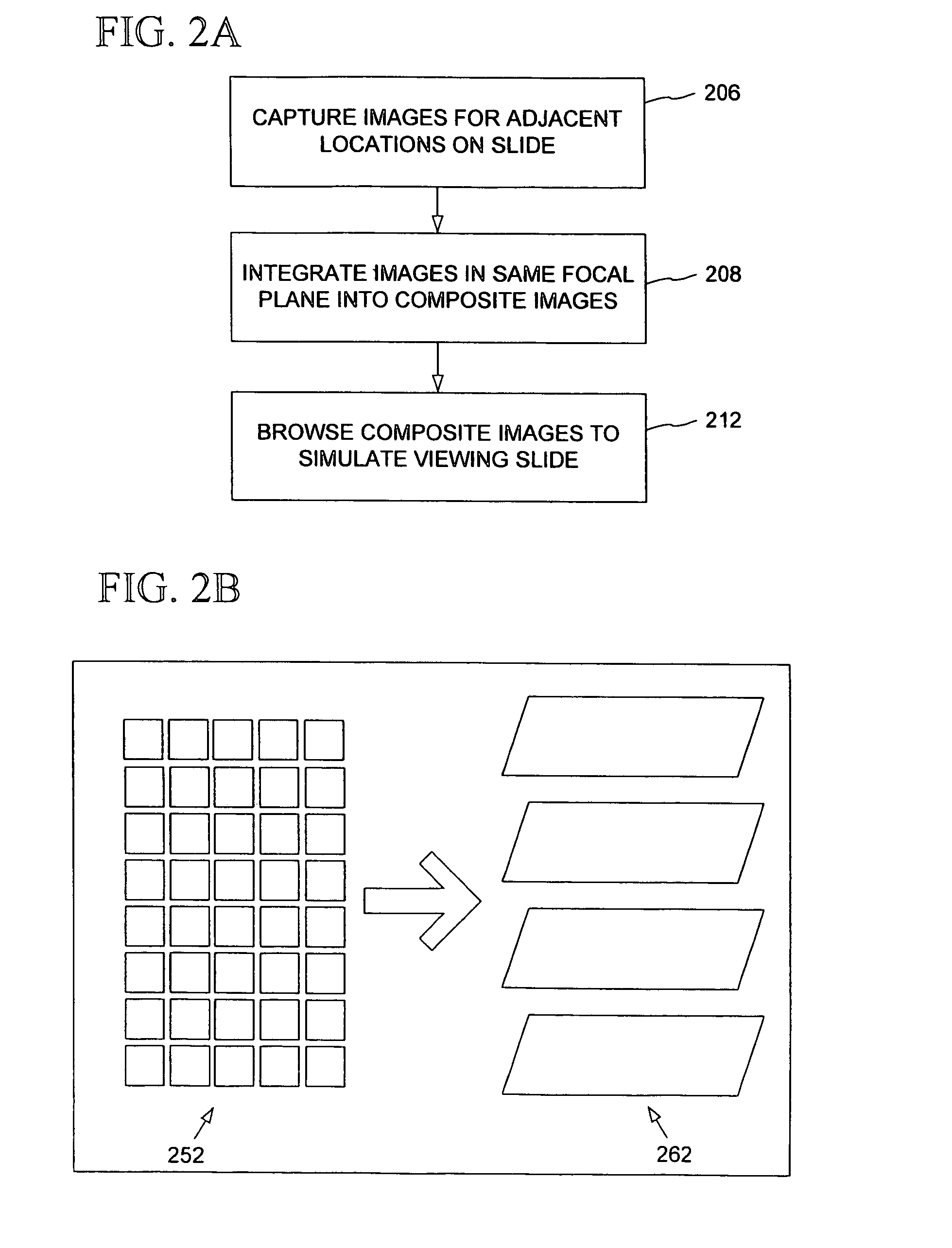
[0103]In an example implementation, images for five focal planes were captured at 40× magnification. There were a total of 18,550 captured images (3,710 per focal plane), each of dimensions 640 by 480 and being about 100 Kb in size. The captured images were saved as separate files according to a file naming convention enabling them to be reloaded for integration. The first letter of the file indicated the focal plane (e.g. A, B, C, D, or E), the next two digits indicated a logical x position, and the next two digits indicated a logical y position.
[0104]After being stitched together, recompressed, and color corrected, a five composite images were generated having a total of 9,976 image portions (including one color-correction image) of dimensions 640 by 480, taking up a total of 636 Mb. These images were saved in a single file, including header information indicating the focal plane and position of each image. 10× composite images (having 108 image portions per composite image) were ...
PUM
 Abstract
Description
Claims
Application Information