Methods and compositions of monoclonal antibodies specific for beta-amyloid proteins

a technology of monoclonal antibodies and beta-amyloid proteins, applied in the field of compositions and compositions of monoclonal antibodies to beta-amyloid proteins, can solve the problems of difficult diagnosis of alzheimer's disease before death, difficult to develop drug therapies, or to treat, and methods for diagnosing ad have not been proven to detect ad in all patients. , to achieve the effect of reducing apoptotic cell death, reducing apoptoti

Inactive Publication Date: 2006-06-13
DIAGENICS INT CORP
View PDF6 Cites 48 Cited by
  • Summary
  • Abstract
  • Description
  • Claims
  • Application Information

AI Technical Summary

Benefits of technology

[0104]The interaction of Aβ1-42 fibers with R7CN was further investigated by cellular methods. In PC12 cells, preaggregated Aβ1-42 (final concentration 0.5 mg / l) induced apoptotic cell death after 24 hrs (FIG. 9A). Necrotic cell death could be excluded, by a LDH assay which revealed that lysis of cell membrane occurred only after 48 hrs (data not shown). Preincubation of aggregated Aβ1-42 with R7CN at a molar ratio of mAb / Aβ from 1:100 reduced apoptotic cell death in PC12 cells by at least 60% (FIG. 9A).
[0105]Aβ peptides induce an inhibition of MTT reduction. The MTT Cell Proliferation Assay is a colorimetric assay system which measures the reduction of a tetrazolium component (MTT) into an insoluble formazan product by the mitochondria of viable cells. After incubation of the cells with the MTT reagent for approximately 2 to 4 hours, a detergent solution is added to lyse the cells and solubilize the colored crystals. The samples are read using an ELISA plate reader at a wavelength of 570 nm. The amount of color produced is directly proportional to the number of viable cells. his assay is an indicator of the mitochondrial activity of living cells and is a widely used as an index of cytotoxicity. Furthermore, this assay seems to be very sensitive to the neurotoxic effects of Aβ as it has been shown in cell culture experiments after exposure to Aβ fragments already at very low concentrations (Leutz et al., J Mol Neurosci 2002 June; 18(3):189–201; Shearman et al., Proc Natl Acad Sci USA Feb. 15, 1994; 91(4):1470–4).
[0106]At a final concentration of 0.1 μmol / l aggregated Aβ1-42 decreased the ability of PC12 cells to reduce MTT by about 60% (FIG. 9B). Incubation of aggregated Aβ1-42 with R7CN prevented the Aβ1-42-induced collapse in the metabolic activity of PC12 cells (FIG. 9B). The most protective effects have been shown with a mixture of Aβ1-42 / R7CN that has been generated by incubation of aggregated Aβ1-42 with R7CN at a mAb / Aβ ratio from 1:500 up to 1:2000 (FIG. 10B). Incubation with an irrelevant monoclonal antibody had no protective effect, either against Aβ1-42 induced apoptosis or on the reduction of MTT. The treatment of aggregated Aβ1-42 with anti-Aβ-mAb R7CN markedly reduced Aβ neurotoxicity in vitro. These processes might also contribute to the reduction of Aβ burden in transgenic mice after vaccination.

Problems solved by technology

Yet, because the presence of the disease is difficult to diagnose without dangerous brain biopsy, the time of onset is unknown.
It is very difficult to diagnose Alzheimer's disease before death, to develop drug therapies, or to treat AD.
But these methods for diagnosing AD have not been proven to detect AD in all patients, particularly at early stages of the disease.
They are also relatively invasive, requiring a spinal tap.
These peptides, however, do not normally cross the blood-brain barrier in amounts necessary for imaging in vivo.
But Congo red is probably not suitable for in vivo diagnosis of β-amyloid deposits in the brain because only 0.03% of an injected dose of iodinated Congo red can enter the brain parenchyma.
Many compounds contain strongly acidic sulfonic acid groups which severely limit entry of these compounds into the brain.
The inability to assess amyloid deposition in AD until after death impedes the study of this devastating illness and researchers' ability to develop effective therapies targeted at preventing or reversing β-amyloid deposition.

Method used


Image

  • Methods and compositions of monoclonal antibodies specific for beta-amyloid proteins
  • Methods and compositions of monoclonal antibodies specific for beta-amyloid proteins
  • Methods and compositions of monoclonal antibodies specific for beta-amyloid proteins

Examples


example 1

Preparation of Aβ1-16 Antigen

[0078]A first step in making an antigenic component comprises synthesis of tetrapalmitoyl-tetrakis-lysine-Aβ1-16-peptide. Two sequential ε-palmitoylated lysines were introduced at the peptide's C-terminus. The first ε-palmitoylated lysine was anchored onto the 4-alkoxylbenzyl alcohol resin by reacting 335 mg of resin with 388 mg of FmocLys(Pal)OH in the presence of DCC (185 mg) and DMAP (20 mg) in dry, freshly distilled dichloromethane according to Merrifield Lu, G., Mojsov, S., Tam, J. P. & Merrified, R. B. (1981) J. Org. Chem. 46, 3433–3436.

[0079]After stirring for three hours at room temperature, the reaction mixture was filtered and thoroughly washed ten times with dry methylene chloride. In order to ensure a complete reaction, the obtained resin was reacted a second time with a fresh portion of FmocLys(Pal)OH, in the presence of DCC and DMAP in dry methylene chloride. The resin was then suspended in 45 ml dry methylene chloride and 5 ml acetic anhyd...

example 2

Preparation of Monoclonal Antibodies to Aβ1-16

[0083]Using tetrapalmitoyl-tetrakis-lysine-Aβ1-16 reconstituted in liposomes with lipid A as an antigen, as made above in Example 1, C57B1 / 6 mice were inoculated i.p. The mice received 5 immunizations over a period of 10 weeks and anti-amyloid antibodies of 1:5000–1:10,000 were measured by ELISA at the end of this interval. See Schenk et al., Nature (London) 400, 173–177 (1999).

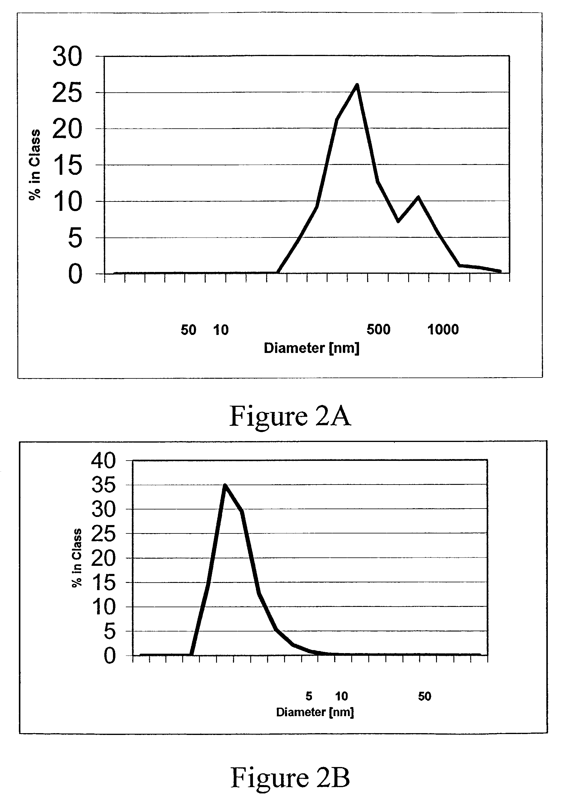
[0084]The mice were tested using the ELISA described below for reaction to synthetic Aβ1-42 fibers. Synthetic, human Aβ1-42 sequence was incubated for seven days in PBS at pH=7.3. This resulted in the formation of fibers, which bind Thioflavin-T, a fluorescent dye emitting at 482 nm.

[0085]To perform the ELISA, microtiter plates were coated with 50 μl of Aβ1-42 solution (1 mg Aβ1-42 / 5 ml PBS) and left overnight at 4° C. Wells were blocked with 200 μl BSA / PBS (0.5% BSA) for two hours at 37° C. and washed with 200 μl of PBS / 0.005% Triton X-100. Dilutions of sera (1:...

example 3

Measurement of Binding to Aβ1-42

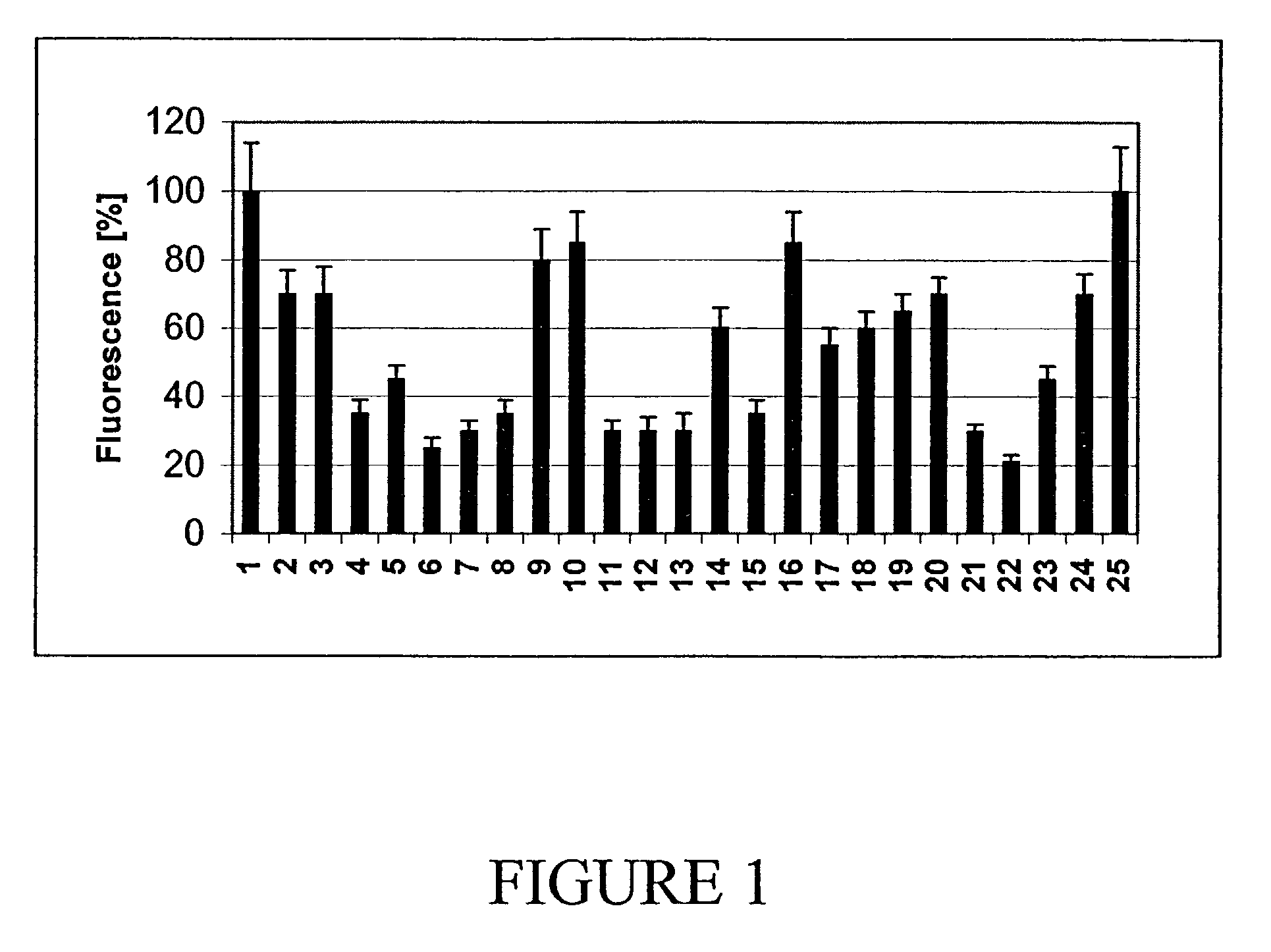
[0090]ELISAs were performed with supernatants of hybridomas from three selected populations (AN-15E6, AN6D4 and AN9C2). The supernatants from these populations showed a significant binding to Aβ1-42 in the assay, and the capability of solubilizing Aβ1-42 fibers in vitro. Subclones were obtained and tested for specific antibodies in an ELISA. The ELISA values of the supernatants of these clones at OD405 are listed in Table 1.

[0091]

TABLE 1ELISA Response of the Supernatants of Hybridoma SubclonesELISANo.Clone Name[OD405] 2AN-15E6-A90.117 3AN-15E6-B90.118 4AN-15E6-C80.159 5AN-15E6-C90.128 6AN-15E6-E80.112 7AN-6D4-C1*0.339 8AN-6D4-C30.298 9AN-6D4-D20.39210AN-6D4-D10.37311AN-6D4-D30.24112AN-6D4-D60.36513AN-6D4-E10.18214AN-6D4-E20.19415AN-6D4-E30.38616AN-6D4-F10.36617AN-6D4-F20.37518AN-6D4-F40.37319AN-9C2C4-E60.14320AN-9C2C4-G60.36521AN-9C2C4-G70.222AN-9C2C3-C110.16623AN-9C2C3-E40.10124AN-9C2C3-G40.168ControlsDMEM / 10% FCS0.124DMEM / 10% FCS / 2% HCF0.113serum f...


PUM

PropertyMeasurementUnit
pHaaaaaaaaaa
concentrationaaaaaaaaaa

Abstract

This invention provides methods and compositions for the detection, diagnosis and treatment of amyloid-associated diseases, in particular, diseases comprising deposition of amyloid assemblies, fibrils, filaments, tangles, or plaques. A preferred composition comprises monoclonal antibodies that specifically bind amyloid proteins, peptides or fragments and change the conformation.

Description

RELATED APPLICATIONS[0001]This application claims benefit of U.S. Provisional Application No. 60 / 336,514 filed Nov. 2, 2001, which is herein incorporated in its entirety.FIELD OF THE INVENTION[0002]The present invention relates to compositions and methods for the diagnosis and treatment of diseases, particularly amyloid-associated diseases. In particular, the invention comprises monoclonal antibodies to beta-amyloid protein that are capable of changing the conformation of the amyloid protein.BACKGROUND OF THE INVENTION[0003]Amyloid protein deposition is associated with cognitive decline in millions of Americans. Amyloid proteins can be deposited in extracellular neufibrillary tangles, within classical or diffuse senile plaques, and in vessel walls. Amyloid plaques in the brain are predominantly composed of (beta)β-amyloid, a 4 kD of protein, 39–43 residues. Beta-amyloid is expressed by a gene located on chromosome 21 and is derived by proteolytic cleavage of a much larger (770 resid...

Claims


Application Information

Patent Timeline
Patent Type & Authority Patents(United States)
IPC IPC(8): A61K39/395C12P21/08C07K16/18
CPCC07K16/18A61K2039/505
Inventor NICOLAU, YVES CLAUDEGREFERATH, RUTH
Owner DIAGENICS INT CORP
Why Eureka
Try Eureka