

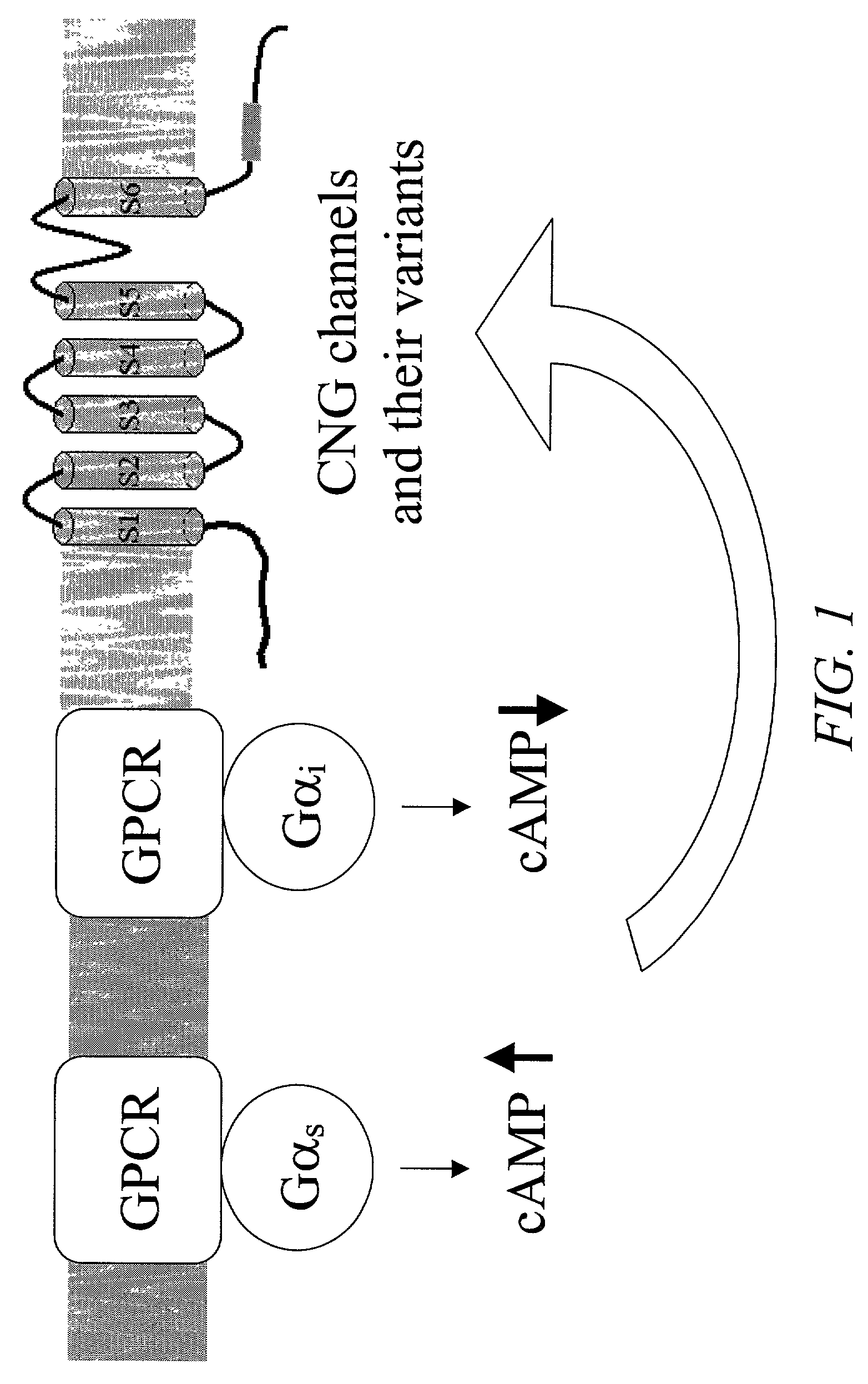
Cell-based assays for G-protein-coupled receptor-mediated activities
a gprotein-coupled receptor and assay technology, applied in the field of cell-based assays for gprotein-coupled receptor-mediated activities, can solve the problems of limited expression and tissue specificity of any individual receptor, inability to probe cells, and inability to meet high throughput screening formats
- Summary
- Abstract
- Description
- Claims
- Application Information
AI Technical Summary
Benefits of technology
Problems solved by technology
Method used
Image

Examples
example 1
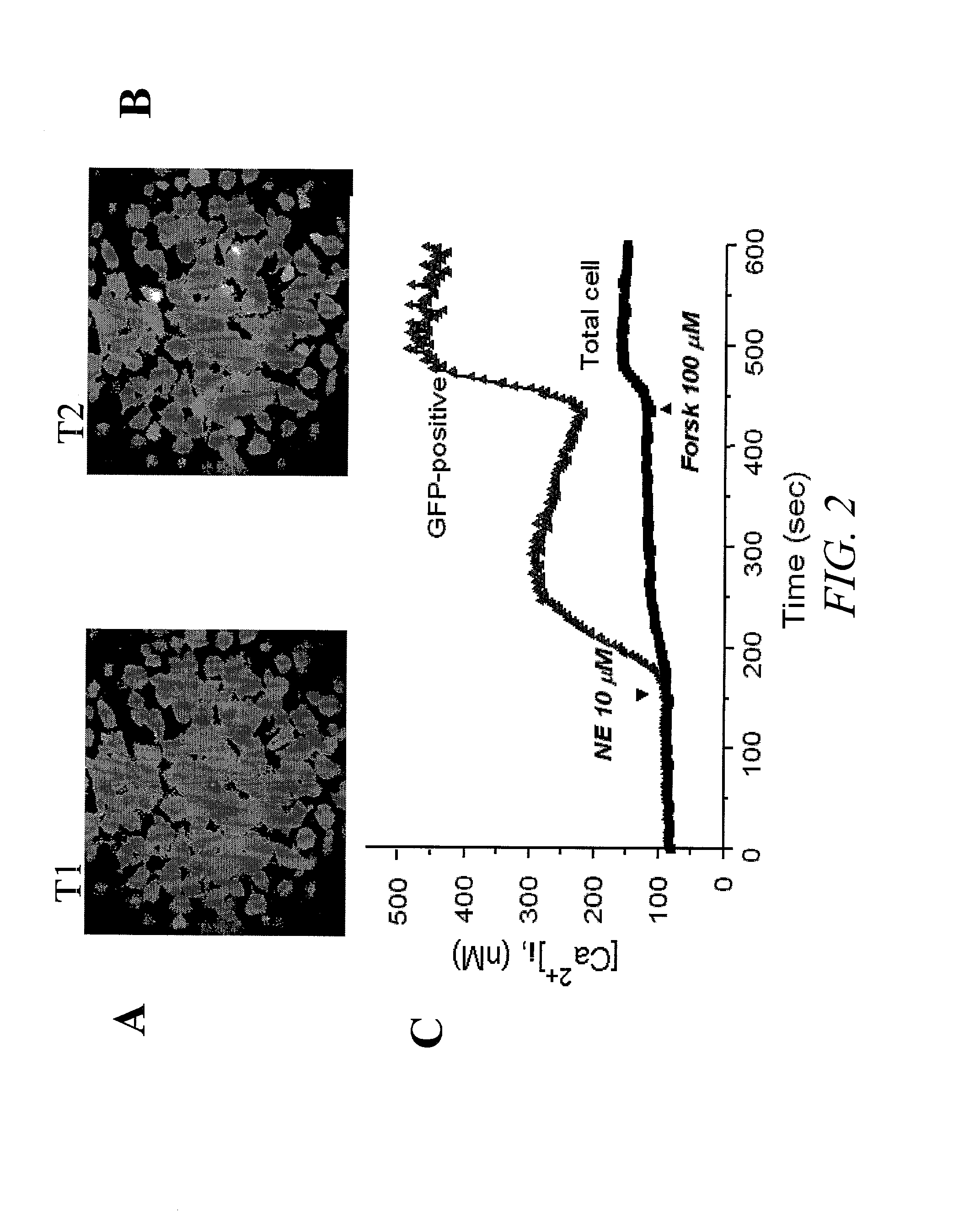
[0131]FIG. 2 shows the results of a single cell imaging assay. In this example, HEK293 cells transiently transfected with a CNG channel (SEQ ID NO:3) for two days were loaded with a calcium fluorescent dye prior to the recordings. Specifically, cells were cultured on a microscope Fisher brand cover glass #1 pre-coated with MATRIGEL (Becton Dickinson, San Jose, Calif.) incubated in culture medium (DMEM with 10% fetal bovine serum) containing 5 μM fura-2 AM (Molecular Probes, Sunnyvale, Calif.) for 0.5 hour at 37° C. Calcium fluorescence recordings were made on Attofluor® RatioVision®, a real-time fluorescence imaging device (ATTO, Rockville, Md.). This system is capable of performing experiments using multiple fluorescent probes such as Fura-2 (for calcium) combined with GFP (transfection marker) in the same cells over the same period of time. Ratio images and graphical data from multiple dyes are displayed on line. This example demonstrates that activation o...
example 2
Comparison of Calcium Sensitive Dyes and Voltage Sensitive Dyes in a Multiwell Format
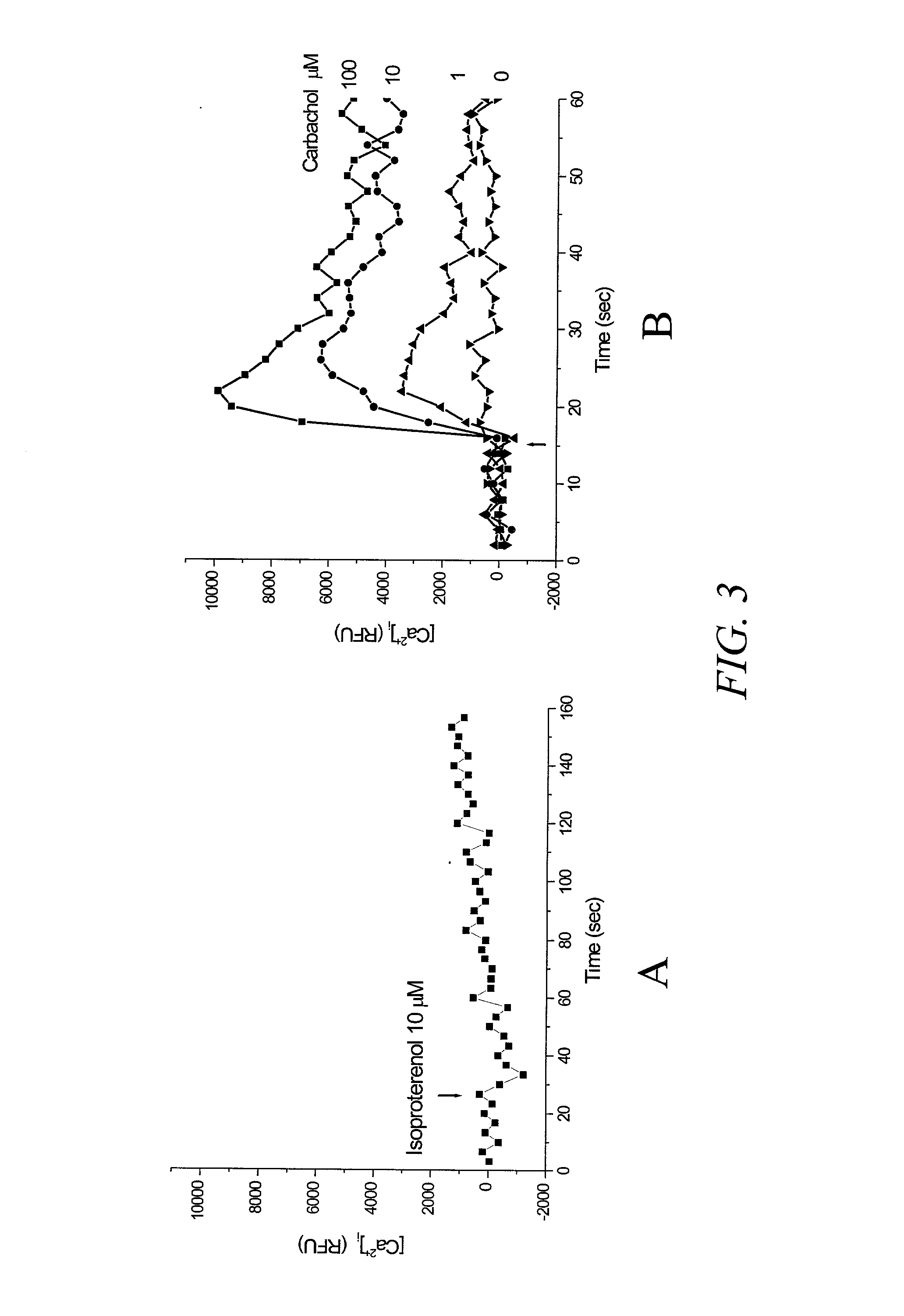
[0132]In this example, recordings were made using a microplate reader FLEXstation (Molecular Devices, Sunnyvale, Calif.) and the protocols provided with the assay kit were adopted for both the calcium assay and the membrane potential assay. FIG. 3 shows the results of a multiwell assay using a mutated CNG channel (SEQ ID NO 5), which is reported to have a lower value EC50 value for cAMP (Rich, et al. 2001, J. Gen. Phys. 118:63–77) than that of SEQ ID NO:3, and therefore expected to be more sensitive to cAMP change. FIG. 3A shows the response to isoproterenol (agonist of β2 adrenergic receptor) determined using a Ca2+ sensitive dye, fluo-4 in cells transiently transfected with the CNG channel. There was no significant change in fluorescence of the dye after stimulating cells with a saturating dose of isoproterenol of 10 μM. FIG. 3B shows the results of activation with carbachol, a muscarinic receptor...
example 3
Assay for Intracellular cAMP in Response to GPCR Activation Using Mutant CNG Channel with a Membrane Potential Dye
[0134]FIG. 5 shows the results of a similar assay in which the voltage-sensitive dye of the membrane potential assay kit was used (Molecular Probes, Sunnyvale, Calif.). DPBS (divalent-free Dulbecco's Phosphate Buffer Salts) supplemented with 0.1 mM MgCl2, 1 mM EGTA and titrated to pH 7.3 was used to reconstitute the voltage-sensitive dye instead of the buffer solution supplied in the commercial kit. HEK293 cells transiently transfected with a CNG channel (SEQ ID NO 5) were loaded with the voltage-sensitive dye at room temperature for about 0.5 hour. A readily detectable change in fluorescence signal was seen at concentrations as low as 0.1 μM isoproterenol that activate β adrenoceptor, as compared to no detectable change with 10 μM isoproterenol using calcium-sensitive dye shown in FIG. 3A. Similarly, FIG. 6 shows the results with membrane potential dye with a different ...
PUM
| Property | Measurement | Unit |
|---|---|---|
| Electric potential / voltage | aaaaa | aaaaa |
| Sensitivity | aaaaa | aaaaa |
Abstract
Description
Claims
Application Information