
Electrosurgical method and apparatus

a surgical method and electrosurgical technology, applied in the field of electrosurgical system, can solve the problems of tissue shrinkage, relatively slow process, etc., and achieve the effect of not producing the expected charring and desiccation of tissue, and considerable effect on treatment tim

Active Publication Date: 2006-11-07
GYRUS ENT
View PDF8 Cites 1398 Cited by
  • Summary
  • Abstract
  • Description
  • Claims
  • Application Information

AI Technical Summary

Benefits of technology

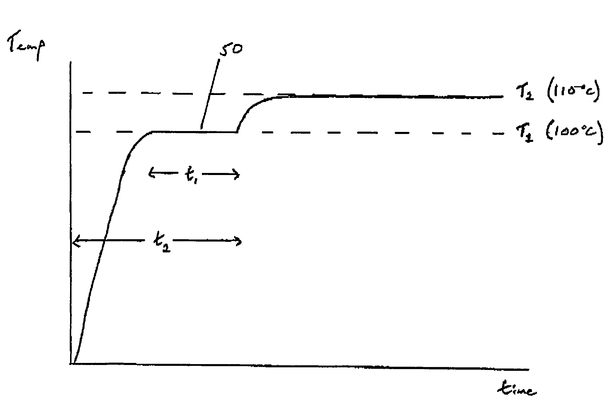
[0014]The control means is adapted to control the generation means such that there is an equilibration phase between the ramp-up phase and the steady state phase. In this equilibration phase, the temperature of the probe is held substantially constant for a period of time at an equilibration temperature to allow the temperature of different parts of the probe to equilibrate. The equilibration temperature is preferably between 90° C. and 105° C., and is typically substantially 100° C. This equilibration phase ensures that all parts of the probe are substantially at the same temperature, and eliminates “hot spots” which are actually at a higher temperature than that detected by the at least one temperature sensor. After the equilibration phase the temperature of the probe is raised to its final steady state operating temperature of between 100° C. and 115° C., preferably substantially 110° C. This equilibration process provides improved temperature control of the probe, and allows temperatures of between 100° C. and 115° C. to be used without the risk that some parts of the probe may be hot enough to cause charring of tissue.
[0020]The present invention is based on the surprising discovery that probe temperatures of slightly higher than 100° C. can produce effective lesions without producing the expected charring and desiccation of the tissue. As stated above, this is contrary to what has been a widespread prejudice against this practice in the field over many years. The explanation for this surprising discovery is not fully understood, but is believed to be based on the phenomenon that the heating of the tissue causes an increase in the concentration of the electrolyte in the tissue in the region of the probe. This causes the boiling point of the electrolyte to be increased, with the result that temperatures of up to 115° C. can be reached without causing charring or significant desiccation of the tissue. This increase in temperature has a considerable effect on treatment time, and it has been found that an acceptable lesion can be produced in around 60 seconds, as opposed to treatment times of 4 to 5 minutes with probes operated at only 85° C.

Problems solved by technology

The lesion is subsequently absorbed by the body with the result that tissue shrinkage is seen to occur.
It is well known that to produce a lesion the temperature of the tissue should be kept below 100° C. Temperatures above 100° C. are known to cause charring and desiccation of the tissue (which can be undesirable as the desiccated tissue is unable to absorb any further RF energy).
The problem with this requirement to maintain the probe temperature below 100° C. is that this can lead to a relatively slow process, requiring anything up to several minutes of treatment time in order to generate the lesion.

Method used


Image

  • Electrosurgical method and apparatus
  • Electrosurgical method and apparatus
  • Electrosurgical method and apparatus

Examples


Embodiment Construction

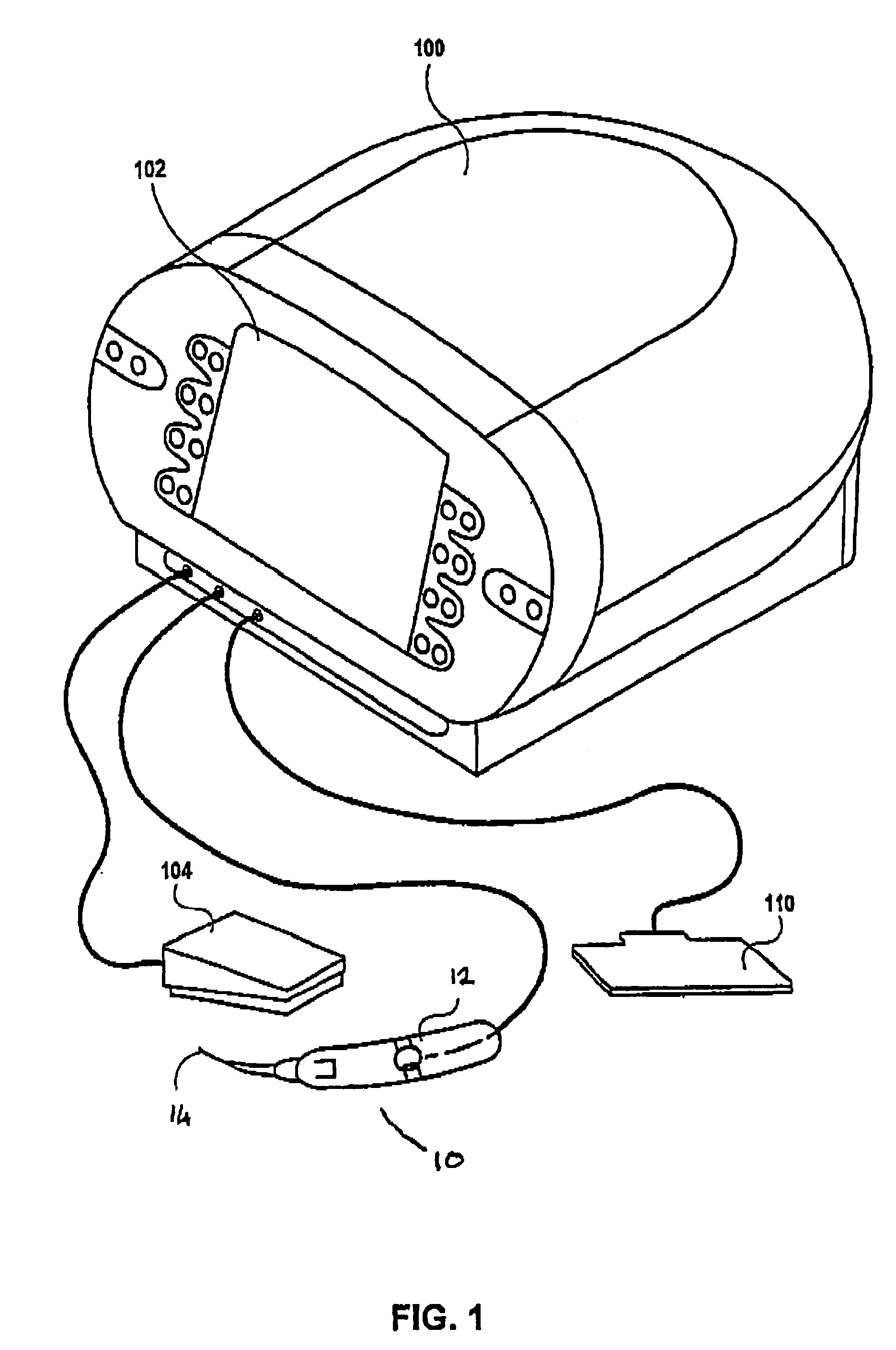
[0027]FIG. 1 shows the apparatus for a typical embodiment of an RF electrosurgical device for forming lesions in body tissue. The system comprises a controller 100 (including an RF power supply) with a user input and display panel 102. Also provided are a foot switch 104, an electrical grounding pad 110 and a probe 10 including a surgical handpiece 12 with a surgical electrode 14. The user input allows the user to input different parameters to affect lesion size, including treatment duration, and total energy delivery.

[0028]The controller 100 converts the low frequency electrical energy supplied by a wall connection (not shown) into the high frequency or RF energy necessary for surgery. The user input and display panel 102 displays relevant parameters and provides buttons and switches for user input to the control systems. The foot switch 104 connected to the controller provides means for switching the unit on and off. The surgical handpiece 12 is also connected to the controller an...


PUM


Abstract

An electrosurgical method and apparatus comprises a probe, at least one temperature sensor, and a controller for generating and controlling electromagnetic energy supplied to the probe. The controller receives signals from the temperature sensor and controls the supply of electromagnetic energy such that the temperature of the probe is ramped up and then maintained at a steady state temperature of between 100° C. and 115° C. In an equilibration phase, between the ramping up and the steady state temperature, the controller holds the temperature of the probe substantially constant for a period of time to allow the temperature of different parts of the probe to equilibrate.

Description

[0001]This nonprovisional application claims the benefit of U.S. Provisional Application No. 60 / 445,455, filed Feb. 7, 2003.FIELD OF THE INVENTION[0002]This invention relates to an electrosurgical system comprising an electrosurgical generator and a handpiece including electrosurgical electrodes. In particular, this invention is directed to electrosurgical systems capable of forming a lesion in body tissue, as is known for the treatment of various medical conditions including airway obstructions and sleep apnea.BACKGROUND OF THE INVENTION[0003]It is well known in the field of electrosurgery that there are two distinct tissue effects which can be achieved by RF, depending on the temperature to which the tissue is raised. The first is the removal of tissue by vaporisation, in which the electrode or electrodes are subjected to relatively high temperatures (typically over 1000° C.). The second is the production of tissue necrosis without the removal of tissue in situ, and it is to this ...

Claims


Application Information

Patent Timeline
IPC IPC(8): A61B19/00A61B18/12A61B18/14
CPCA61B18/1206A61B18/1477A61B2018/00577
Inventor AMOAH, FRANCIS
Owner GYRUS ENT
Learn More