Supercharge Your Innovation With Domain-Expert AI Agents!

Methods of assaying receptor activity

a technology of gpcr and activity, which is applied in the field of methods of detecting gprotein coupled receptor activity, can solve the problems that the common role of -arrestin in gpcr desensitization has not been established

Inactive Publication Date: 2006-11-21
DUKE UNIV
View PDF114 Cites 16 Cited by
  • Summary
  • Abstract
  • Description
  • Claims
  • Application Information

AI Technical Summary

Benefits of technology

The present invention relates to a method for detecting G protein coupled receptor (GPCR) activity using a β-arrestin protein and a detectable molecule. The detectable molecule can be an optically detectable molecule such as Green Fluorescent Protein (GFP). The method involves expressing a GPCR and a β-arrestin protein in a cell, and then exposing the cell to a known GPCR agonist or a sample solution containing a potential agonist or antagonist. The movement of the detectable molecule from the cell cytosol to the membrane edge after exposure indicates GPCR activity or the absence of activity. The invention can be used to screen for GPCR agonists or antagonists, and to study the function of β-arrestin protein in GPCR signaling.

Problems solved by technology

Moreover, except for a limited number of receptors, a common role for β-arrestin in GPCR desensitization had not been established.

Method used


Image

  • Methods of assaying receptor activity
  • Methods of assaying receptor activity
  • Methods of assaying receptor activity

Examples


example 1

Materials and Methods

[0088]Materials: Isoproterenol was obtained from Sigma RBI. Anti-mouse antibody was obtained from Sigma Chemicals or Molecular Probes. Mouse monoclonal antibody against the 12CA5 epitope was obtained from Boehringer Mannheim. Cell culture media was obtained from Mediatech and fetal bovine serum from Atlanta Biologicals. Physiological buffers were from Gibco-Life Technologies Inc. Restriction enzymes were obtained from Promega or New England Biolabs, T4 ligase was from Promega, and Hot Tub DNA polymerase from Amersham. Commercially available plasmids containing variants of Green Fluorescent Protein were obtained from Clontech.

[0089]Cell Culture and Transfection: HEK-293 and COS cells were maintained and transfected as described by Barak et al., Mol. Pharm. 51:177 (1997). Cells containing both beta2 adrenergic receptor and β-arrestin constructs were transfected with between 5–10 .mu.g of receptor cDNA in pcDNA1 / AMP and 0.5–1 ·mu·g of βarr2-GFP cDNA per 100 mm dish...

example 2

Construction of β-arrestin2-GFP Plasmid

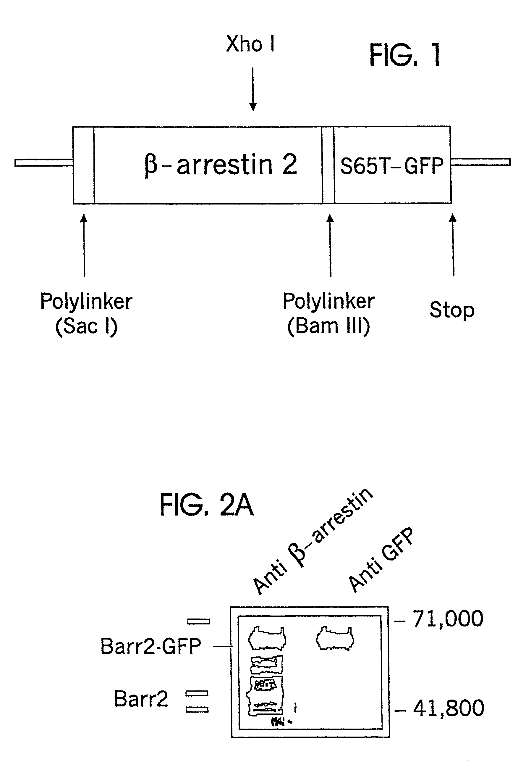
[0092]β-arrestin2 cDNA in the plasmid pCMV5 was used as a template. Oligonucleotide primers surrounding a distal XhoI restriction site and the C-terminal stop codon of β-arrestin2 were used to replace the stop codon with an in frame BamHI restriction site by directed mutagenesis (Valette et al. Nucleic Acids Res. 17:723 (1989); Attramadal et al., J. Biol. Chem. 267:17882 (1992); Lohse et al., Science 248:1547 (1990)). The XhoI, BamHI segment was isolated. This segment was ligated to the N-terminal portion of β-arrestin CDNA (cut from pCMV5 by SacI and XhoI) in the polylinker of a plasmid that had been previously digested with ScaI and BamHI and that contained S65T-Green Fluorescent Protein distal and in frame to the site of β-arrestin cDNA insertion. Lohse et al., Science 248:1547 (1990). The resulting β-arrestin-GFP construct was isolated following insertion and growth in E. coli. Constructs were verified by sequencing.

[0093]A linear model of ...

example 3

Characterization of βarr2-GFP Expressed by HEK-293 Cells

[0094]Homogenates of HEK-293 cells transformed with the plasmid of Example 2 were studied using known Western Blot techniques. The results showed that HEK-293 cells expressed both endogenous β-arrestin and the βarr2-GFP conjugate.

[0095]Western blots of homogenates of HEK-293 cells transfected with the plasmid of Example 2 and expressing βarr2-GFP were performed. An equal amount of homogenate material was loaded into each of two lanes (FIG. 2A). The left lane was exposed to anti-βarrestin antibody (Menard et al., Mol. Pharm. 51:800 (1997)), whereas the right lane was exposed to a mouse monoclonal antibody against GFP. The βarr2-GFP fusion protein is approximately 50% larger than β-arrestin2, and would thus be expected to migrate more slowly than β-arrestin on SDS-Page.

[0096]Exposure to anti-βarrestin antibody revealed multiple bars (left lane); exposure to anti-GFP monoclonal antibody revealed a single bar (right lane). The posi...


PUM

PropertyMeasurementUnit
temperaturesaaaaaaaaaa
fluorescentaaaaaaaaaa
Green Fluorescentaaaaaaaaaa

Abstract

Described are methods of detecting G-protein coupled receptor (GPCR) activity in vivo and in vitro; methods of assaying GPCR activity; and methods of screening for GPCR ligands, G Protein-coupled receptor kinase (GRK) activity, and compounds that interact with components of the GPCR regulatory process.

Description

[0001]This application is a continuation of U.S. patent application Ser. No. 09 / 631,468, filed Aug. 3, 2000, now abandoned, which is a continuation of U.S. patent application Ser. No. 09 / 233,530, filed Jan. 20, 1999, now issued as U.S. Pat. No. 6,110,693, which is a continuation of U.S. patent application Ser. No. 08,869,568, filed Jun. 5, 1997, now issued as U.S. Pat. No. 5,891,646, the disclosure of which is incorporated by reference in its entirety.FEDERALLY SPONSORED RESEARCH[0002]This invention was made with Government support under National Institutes of Health Grant No. HLO3422-02 and NS 19576. The Government has certain rights to this invention.FIELD OF THE INVENTION[0003]This invention relates to methods of detecting G-protein coupled receptor (GPCR) activity in vivo and in vitro, and provides methods of assaying GPCR activity, and methods of screening for GPCR ligands, G protein-coupled receptor kinase (GRK) activity, and compounds that interact with components of the GPCR...

Claims


Application Information

Patent Timeline
Patent Type & Authority Patents(United States)
IPC IPC(8): G01N33/566G01N21/76G01N33/53C07K14/47C12N15/62
CPCC07K14/47C12N15/62G01N33/5008C07K2319/00C07K2319/60G01N2333/726G01N2333/91
Inventor BARAK, LAWRENCE S.CARON, MARC G.FERGUSON, STEPHEN S.ZHANG, JIE
Owner DUKE UNIV
Features
  • R&D
  • Intellectual Property
  • Life Sciences
  • Materials
  • Tech Scout
  • Unparalleled Data Quality
  • Higher Quality Content
  • 60% Fewer Hallucinations
Learn More