System and method for mining quantitive information from medical images

a quantitive information and medical image technology, applied in the field of medical imaging, can solve the problems of inability to accurately and accurately describe the shape and size information, the method is severely disadvantageous in the cost and time taken to extract the size and shape information, and the manual tracking of the shape and size in serial data is extremely difficult to do manually. , to achieve the effect of enhancing clinical diagnosis and research

Active Publication Date: 2007-01-02
CLOUD SOFTWARE GRP INC
View PDF2 Cites 131 Cited by
  • Summary
  • Abstract
  • Description
  • Claims
  • Application Information

AI Technical Summary

Benefits of technology

[0013]The present invention comprises an Imaging System for quantitative information extraction from multi-modal medical images such as CT, MR, ultrasound and PET, for tracking of this information over time, and for archiving and mining of this information that overcome the deficiencies of the prior art. Furthermore, the system uniquely combines various image segmentation methods, image registration methods, image feature extraction methods, audit trail recording, and database storage and retrieval methods and routines into seamless environment capable of enhancing clinical diagnosis and research.

Problems solved by technology

Both these currently practiced methods suffer from lack of accuracy and precision (due to inter-observer variability.)
In addition, the latter method has a severe disadvantage in terms of the cost and time taken to extract size and shape information.
Moreover, manual tracking of shapes and sizes in serial data is extremely difficult to do manually.
The conventional methods are deficient in that they do not allow the combination of various segmentation methods, registration methods, feature extraction methods, and database storage and retrieval.
The conventional methods are further deficient in that features that have been extracted cannot be saved back into the database, linked, quantified and queried upon.
Based upon these deficiencies alone none of these systems can perform a dual purpose as an image extracting and image mining system.
The prior art is also deficient in that prior art systems do not allow quantitative tracking of shapes and sizes of anatomy or pathology over time.
Furthermore, the prior art systems are deficient in that they do not provide a fast, automatic, multimodal registration method or process.
The quantitative features extracted are not stored in the database so that they fail to link and quantify the data and do not support the mining of quantitative information.
Moreover, none of these systems support tracking of serial / longitudinal data.
Nor do the prior art systems provide multi-channel tissue classification or shape-based interpolation.
These systems do not provide any level of database or image mining support.

Method used


Image

  • System and method for mining quantitive information from medical images
  • System and method for mining quantitive information from medical images
  • System and method for mining quantitive information from medical images

Examples


Embodiment Construction

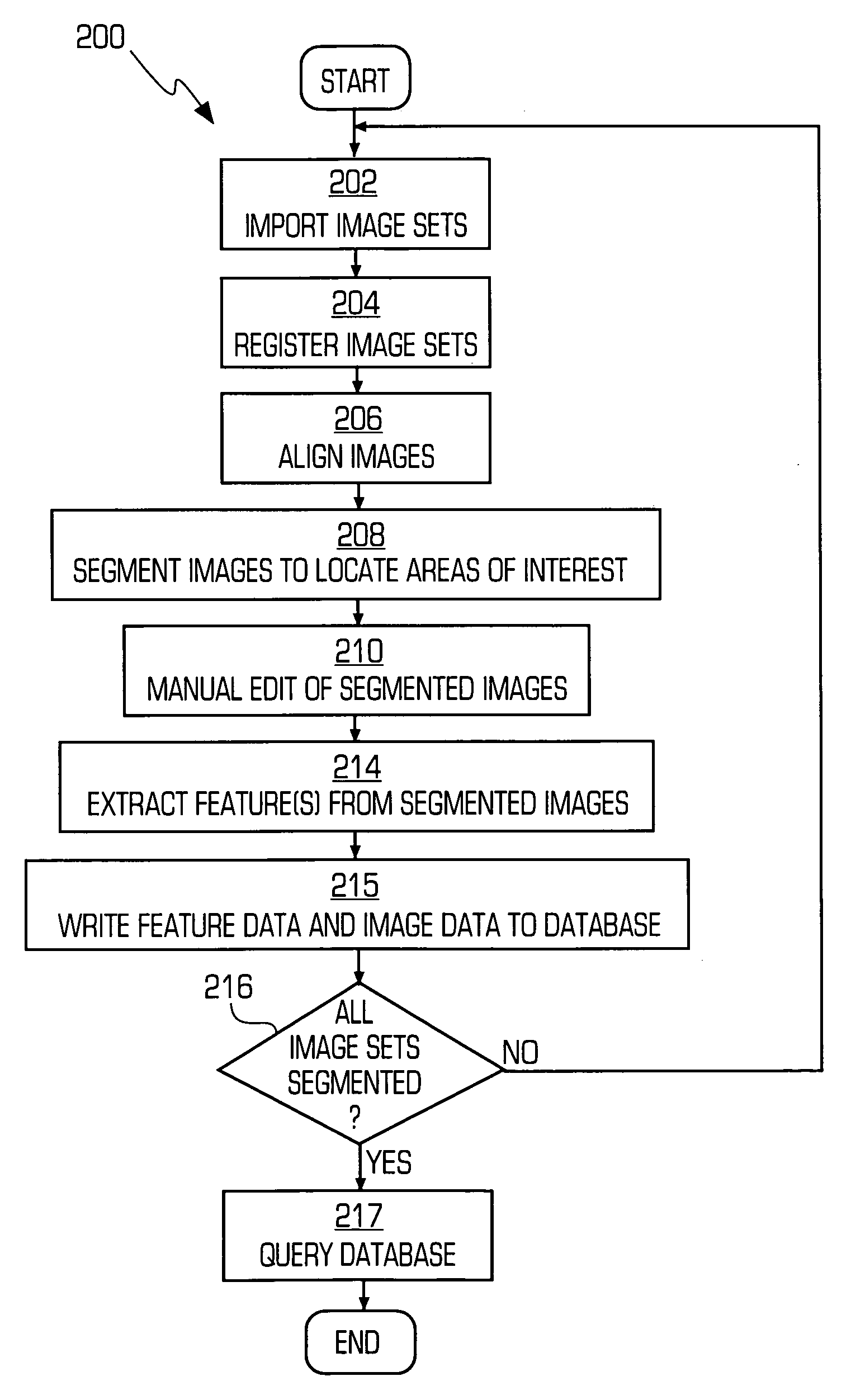
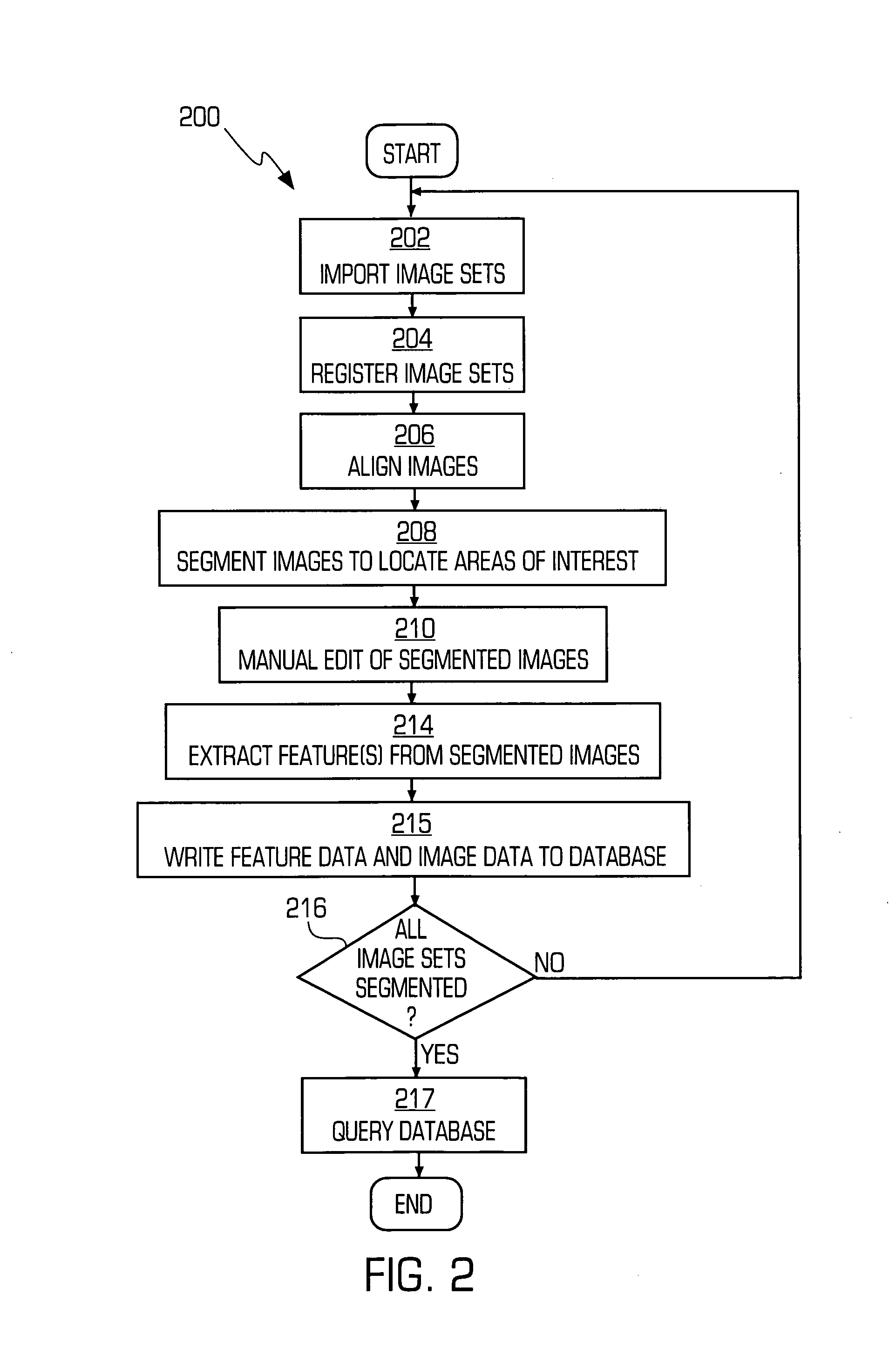
[0047]Reference will now be made in detail to the present embodiments of the invention, examples of which are illustrated in the accompanying drawings. Wherever possible, the same reference numbers will be used throughout the drawings to refer to the same or like parts (elements).

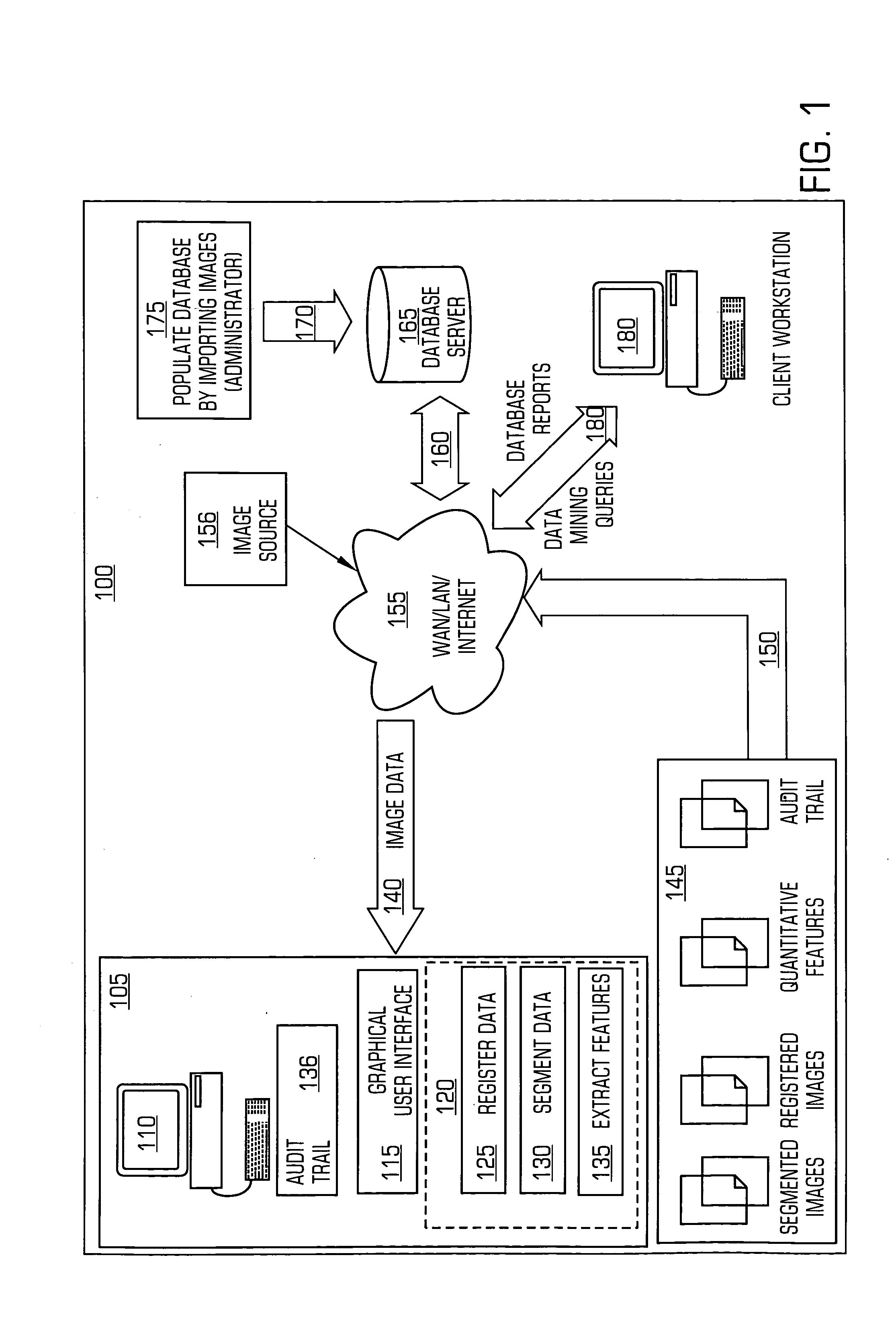
[0048]In accordance with the invention, the present invention can be characterized according to one aspect of the present invention as a system for multi-modality image registration and quantitative feature extraction for multiple patients. The system includes an imaging workstation having a data processor and memory in communication with a database server. The data processor is capable of inputting and outputting data and instructions to peripheral devices and operating pursuant to a software product and accepts instructions from a graphical user interface capable of interfacing with and navigating the imaging software product. The instructions may also be stored and executed from a batch file. The imaging...


PUM


Abstract

A system for image registration and quantitative feature extraction for multiple image sets. The system includes an imaging workstation having a data processor and memory in communication with a database server in communication. The a data processor capable of inputting and outputting data and instructions to peripheral devices and operating pursuant to a software product and accepts instructions from a graphical user interface capable of interfacing with and navigating the imaging software product. The imaging software product is capable of instructing the data processor, to register images, segment images and to extract features from images and provide instructions to store and retrieve one or more registered images, segmented images, quantitative image features and quantitative image data and from the database server.

Description

CROSS REFERENCE TO RELATED APPLICATIONS[0001]This application claims priority under 35 U.S.C. § 119(e) to provisional application No. 60 / 329,843, filed Oct. 15, 2001, entitled “METHOD AND SYSTEM FOR MINING QUANTITIVE INIFORMATON FROM MEDICAL IMAGES”, which is hereby fully incorporated by reference and provisional application No. 60 / 329,827, filed Oct. 15, 2001, entitled “Method and System for Image Analysis and Syntactic Searches”, which is hereby fully incorporated by reference.FIELD OF INVENTION[0002]This invention is directed to the field of medical imaging in general and particularly is directed towards the use of medical imaging in clinical diagnosis, clinical trials and clinical research, including drug discovery and clinical trials of drugs and more particularly focuses on quantitative information extraction from medical images from one or more modalities, tracking of this information over time, and archiving and mining of this information.BACKGROUND OF THE INVENTION[0003]Ext...

Claims


Application Information

Patent Timeline
Patent Type & Authority Patents(United States)
IPC IPC(8): G06K9/32G06K9/00G16Z99/00
CPCG06K9/00G06K2209/05G16H50/70G16Z99/00G06V2201/03G06F18/00
Inventor CHALANA, VIKRAMFOGARASI, STEPHENNG, LYDIAOELUND, JOHNPATHAK, SAYANRACKI, STEVENSPARKS, BOBBIWYMAN, BRADLEY
Owner CLOUD SOFTWARE GRP INC
Why Eureka
Try Eureka