Abdominal organ dynamic contrast enhanced magnetic resonance imaging method based on compressed sensing

A dynamic contrast enhancement, magnetic resonance imaging technology, applied in sensors, instruments, measurement of magnetic variables, etc., can solve the problems of reduced image signal-to-noise ratio, low signal-to-noise ratio, hindered time resolution, etc., to reduce the number of filling points, measure Accurate and shortened acquisition time

Active Publication Date: 2015-10-28
PEKING UNIV
View PDF5 Cites 14 Cited by
  • Summary
  • Abstract
  • Description
  • Claims
  • Application Information

AI Technical Summary

Problems solved by technology

However, the parallel imaging technology leads to a decrease in the signal-to-noise ratio (SNR) of the image, and the acceleration factor is usually limited to less than three times. The higher the acceleration rate, the lower the signal-to-noise ratio, which hinders the further improvement of time resolution.

Method used


Image

  • Abdominal organ dynamic contrast enhanced magnetic resonance imaging method based on compressed sensing
  • Abdominal organ dynamic contrast enhanced magnetic resonance imaging method based on compressed sensing
  • Abdominal organ dynamic contrast enhanced magnetic resonance imaging method based on compressed sensing

Examples


Embodiment Construction

[0017] The present invention is described in detail below in conjunction with the accompanying drawings. However, it should be understood that the accompanying drawings are provided only for better understanding of the present invention, and they should not be construed as limiting the present invention.

[0018] The method for dynamic contrast-enhanced magnetic resonance imaging of abdominal organs based on compressed sensing of the present invention includes pulse sequence generation, data sampling of abdominal organs and image reconstruction. The specific contents are:

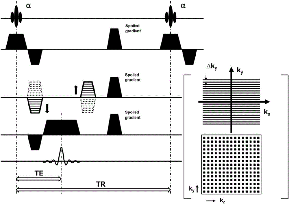
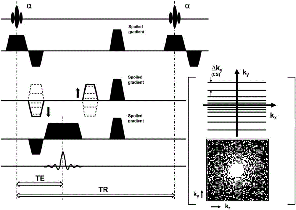
[0019] 1. If figure 2As shown, the MRI pulse sequence is composed of a series of pre-pulses, radio frequency excitation pulses, spatial encoding gradients and signal relaxation. The magnetic resonance imaging pulse sequence of the present invention includes a three-dimensional gradient echo excitation pulse, a spatial encoding gradient and a signal relaxation sequence, which are described respectively bel...


PUM


Abstract

The invention relates to an abdominal organ dynamic contrast enhanced magnetic resonance imaging method based on compressed sensing. The method concretely includes the steps: 1), a magnetic resonance imaging pulse sequence includes a three dimensional gradient echo excitation pulse, a space coding gradient and a signal relaxation sequence, the three of which are explained respectively as follows: 1.1) setting parameters of a radio frequency excitation pulse of a three dimensional gradient echo sequence; 1.2) optimizing a choose-layer phase coding kz and an inner-layer phase coding ky respectively, that is to say, carrying out sub-sampling according to a CS theory, the frequency coding direction kx being fully sampling; and 1.3) applying spoiled gradient to an x gradient direction, a y gradient direction and a z gradient direction in terms of the signal relaxation sequence; 2) a magnetic resonance imaging system carries out compressed sampling of k-space data of all phases of DCE-MRI scanning for an abdominal organ on the basis of a CS optimized magnetic resonance imaging pulse sequence, and obtains original sampling data of a time sequence; and 3) CS reconstruction of the original sampling data is conducted, that is to say, a DICOM image of the abdominal organ is reconstructed and obtained on the basis of a non-linear algorithm with a minimized 1<1> normal form. The method can be widely applied to abdominal organ dynamic contrast enhanced magnetic resonance imaging.

Description

technical field [0001] The invention relates to a dynamic contrast-enhanced magnetic resonance imaging method of abdominal organs based on a compressed sensing (CS) optimized sequence, which can provide dynamic contrast-enhanced images of abdominal organs with high time resolution and high signal-to-noise ratio, and belongs to the technical field of magnetic resonance medical imaging . Background technique [0002] Abdominal organ diseases, such as chronic kidney disease (chronic kidney disease, CKD), have become a global public health problem. In developed countries, the prevalence rate of the general population is as high as 6.5% to 16%, and the incidence rate is increasing year by year. Finances carry a heavy burden. According to the results of the 2012 "Chronic Kidney Disease Epidemiological Survey in China", the prevalence of chronic kidney disease in the adult population in my country has reached 10.8%, and it is estimated that there are 120 million adult chronic kidn...

Claims


Application Information

Patent Timeline
Patent Type & Authority Applications(China)
IPC IPC(8): G01R33/561A61B5/055
Inventor 陈斌张珏王霄英方竞
Owner PEKING UNIV
Why Eureka
Try Eureka