Apparatus and method for the detection and quantification of joint and tissue inflammation

a technology for joint and tissue inflammation and apparatus, applied in the field of medical methods and apparatus, can solve the problems of inability to reliably establish the presence of inflammation, crude and inconsistent assessment, and ultimately no definite correlation, and achieve the effect of convenient us

Active Publication Date: 2007-06-05
ECTOSCAN SYST LLC
View PDF9 Cites 48 Cited by
  • Summary
  • Abstract
  • Description
  • Claims
  • Application Information

AI Technical Summary

Benefits of technology

[0027]Another primary object of this invention is to provide a relatively easy to use apparatus and a method for the detection and quantification of joint and tissue inflammation.

Problems solved by technology

This method of questioning and physical examination of the patient, however, does not reliably establish the presence of inflammation.
Further, this method results in an assessment that is only crudely and inconsistently quantifiable.
Accordingly, such assessments are highly subjective and ultimately have no definite correlation with the actual degree of inflammation.
Even if the physician detects some amount of joint or tissue swelling by palpation, there is no precise and reproducible way of determining whether the swelling represents newly inflamed tissue or just residual thickened and scarred tissue from previous, now quiescent inflammation.
Detection of skin warmth is useful but cannot be quantified by simple palpation.
The determination of tenderness is also useful but it only inconsistently correlates with actual inflammation.
Unfortunately, problems of accuracy and reproducibility arise due to placement and stability of the protractor.
Unfortunately, such tests are nonspecific and indicate changes taking place throughout the patient's body rather than to inflammatory changes taking place in localized areas, such as a knee or finger.
Unfortunately, this method has numerous problems.
Further, as with blood count tests, such methods only indicate the possibility of inflammation and reflect a systemic rather than a local situation.
Like the other blood tests used for detecting inflammation, CRP suffers from the lack of sensitivity, lack of specificity and numerous confounding factors.
As a result of the numerous difficulties associated with the various methods of assessing inflammation using blood tests and since these methods do not include measurement of the five above-mentioned markers of inflammation, such methods are indirect at best and only useful in obtaining adjuncts for detecting and quantifying inflammation.
Methods for detecting inflammation using plain radiographs, such as plain X-rays, have proved to be inadequate It has been found that if a patient has swelling capable of being shown in a X-ray, the swelling level would also be clinically evident on physical examination thereby rendering a X-ray unnecessary.
Such methods, however, are relatively expensive, may be insensitive to very mild degrees of inflammation, and generally do not detect forms of inflammation that involve only minimal or no swelling.
Further, an MRI cannot be used for individuals having pacemakers or metallic implants located near the subject area.
Another problem with using CT and MRI scans is the difficulty of interpreting the results of the scans making them unsuitable for routine outpatient testing in a typical medical office or clinic.
Unfortunately, however, thermography is relatively insensitive to mild degrees of inflammation, it cannot detect any of the signs of inflammation except warmth, it is difficult to reproduce due to changes in regional blood flow, for example caused by emotional states, and it is relatively expensive and technically difficult, requiring the patient to be unclothed and placed in a climate-controlled room where ambient temperature and humidity are assiduously constant and stabilized.

Method used


Image

  • Apparatus and method for the detection and quantification of joint and tissue inflammation
  • Apparatus and method for the detection and quantification of joint and tissue inflammation
  • Apparatus and method for the detection and quantification of joint and tissue inflammation

Examples


Embodiment Construction

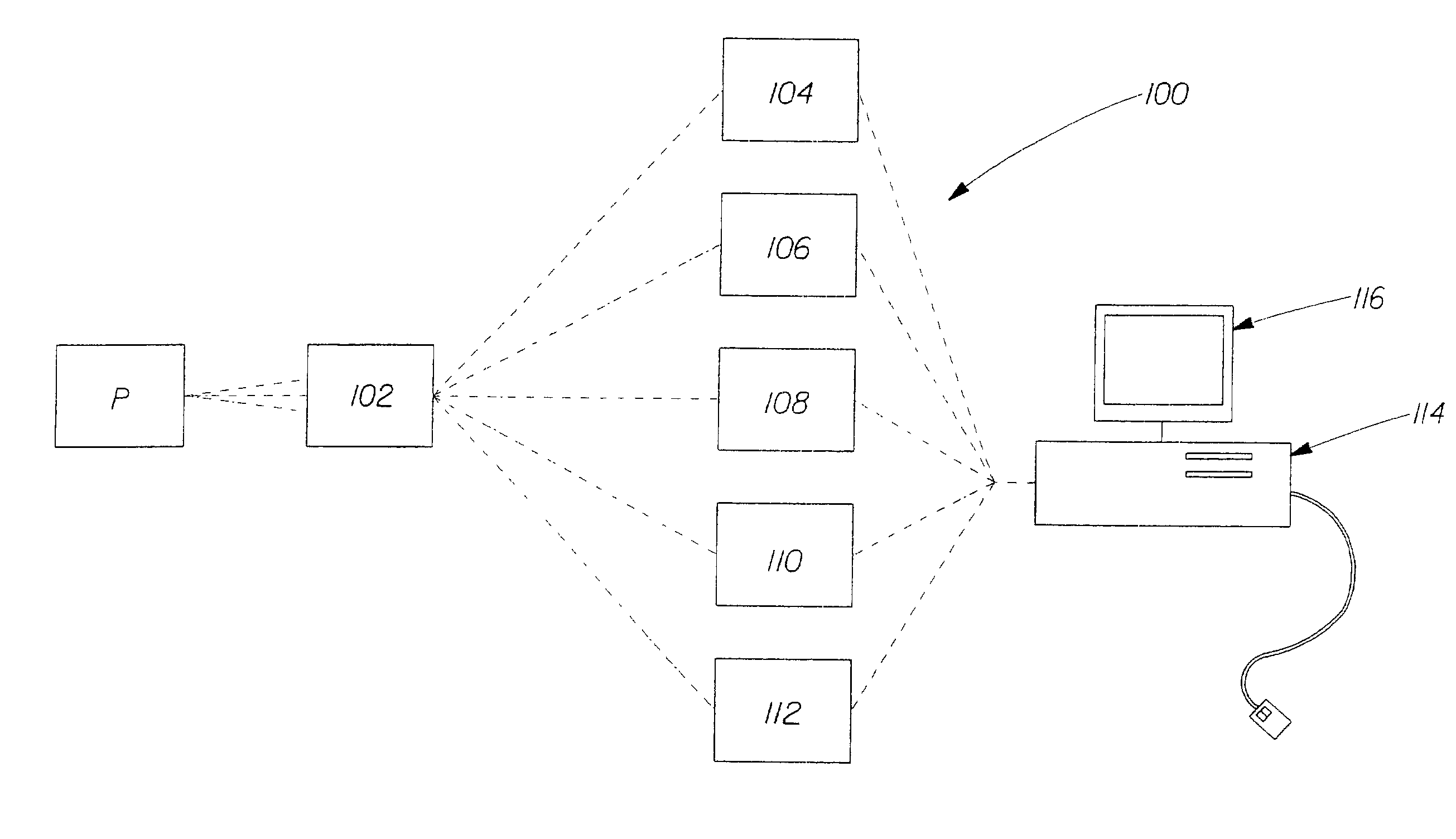
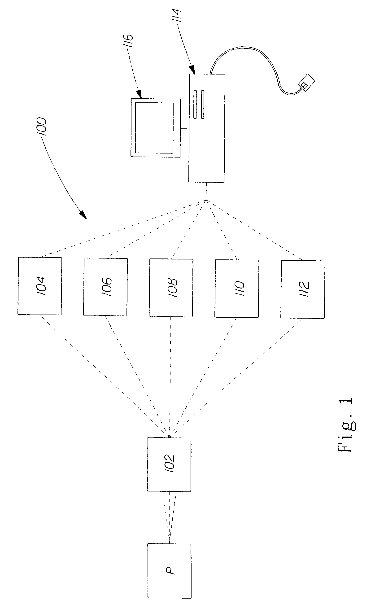
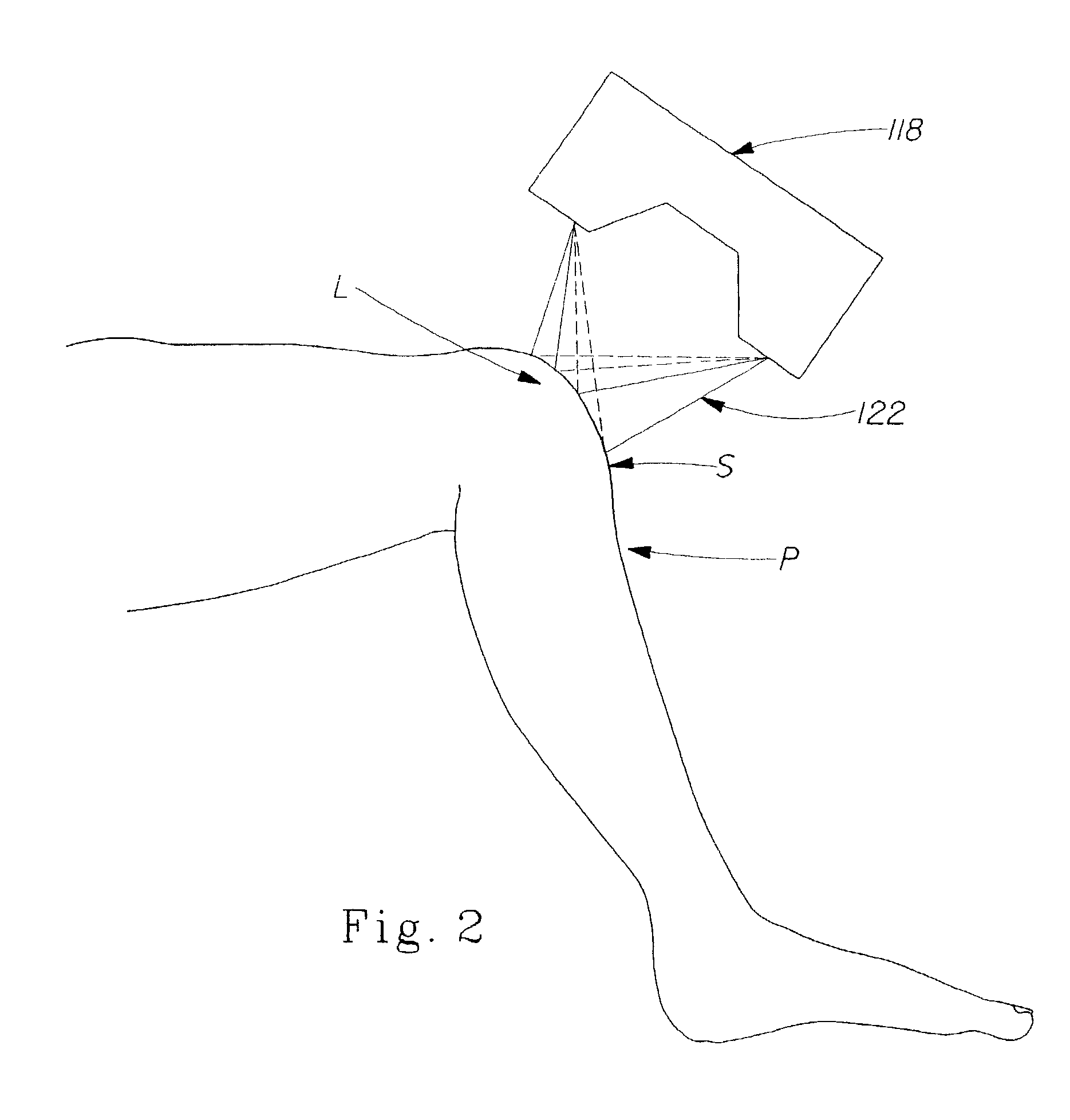
[0040]Referring to FIGS. 1 and 2, a non-human observer or non-clinician apparatus for the detection and quantification of joint and tissue inflammation, generally designated 100, is shown comprising a sensing component 102 for obtaining and collecting data indicative of the surface at the location L of the joint or tissue of a patient P. Preferably, the sensing component 102 comprises various components for assessing inflammation such as a device for detecting swelling 104, such as an optical imaging system, by providing dimensional measurement of the location L of the joint or tissue of the patient P being examined for inflammation, a range-of-motion device 106 for determining the patient's range of motion of the joint being examined, a color analyzing device 108 for analyzing color of the patient's skin at the location L of the joint or tissue, a temperature measuring device 110 for measuring the temperature of the skin S of the patient P at the location L of the joint or tissue, ...


PUM


Abstract

The apparatus and method for the detection and quantification of joint and tissue inflammation (100) of the present invention comprises a sensing component (102) for obtaining and collecting data indicative of the surface or dimensions of the joint or tissue. The sensing component (102) comprises various components for obtaining measurements of the cardinal signs of inflammation. Preferably the sensing component (102) includes a device for detecting swelling (104), a color analyzing device (108), a temperature measuring device (110), a pain or tenderness detection device (112), an archival and retrieval device (114), and a display device (116). In operation a patient is placed in position for examination of the joint or tissue area measurements are taken of one or more of the cardinal signs of inflammation at the joint or tissue area, the measurements are automatically stored in a archival storage and retrieval device (114), and from the measurements an inflammation score is derived which can be compared against a baseline.

Description

TECHNICAL FIELD[0001]The subject invention relates to medical method and apparatus and more particularly to apparatus and method for the detection and quantification of joint and tissue inflammation.BACKGROUND OF THE INVENTION[0002]Most people by age 50 develop some degree of arthritis. Usually, this takes the form of degenerative joint disease, or osteoarthritis that eventually afflicts nearly everyone by the time they are 65 years old. Another form of arthritis, rheumatoid arthritis, affects approximately 1% of the population with a prevalence that approaches 2% of males and 5% of females by age 65. When lupus, gout, infectious processes, metabolic processes, toxins, cancers, and the more than 100 other types of arthritis are added to these numbers, nearly everyone, if they live long enough, will suffer joint pain and arthritis. The ultimate prevalence of joint diseases is further increased by fractures, athletic injuries, neuromuscular disorders, and congenital deformities.[0003]...

Claims


Application Information

Patent Timeline
Patent Type & Authority Patents(United States)
IPC IPC(8): A61B5/117A61B5/103A61B5/00A61B5/107
CPCA61B5/103A61B5/411A61B5/4528A61B5/4824A61B5/01A61B5/107
Inventor THOMSON, PAUL E.
Owner ECTOSCAN SYST LLC
Why Eureka
Try Eureka