Vision catheter system including movable scanning plate

a scanning plate and catheter technology, applied in the field of medical devices, can solve the problems of large size and stiffness of the endoscope, difficult assembly, and difficult assembly of the endoscope, and achieve the effects of easy assembly, imaging capabilities, and relatively inexpensive and disposabl

Inactive Publication Date: 2007-09-25
BOSTON SCI SCIMED INC
View PDF20 Cites 88 Cited by
  • Summary
  • Abstract
  • Description
  • Claims
  • Application Information

AI Technical Summary

Benefits of technology

[0006]The present invention is directed to a vision catheter system. In accordance with one aspect of the invention, the device is in the form of a disposable imaging catheter that produces high-resolution color images that are comparable to those obtained from an endoscope. The device may also function as a guidewire to guide larger devices to areas of interest to facilitate diagnosis and treatment within various lumens of the human body, such as the vasculature, GI, urology (URO) and biliary tracts. The vision catheter system of the present invention is intended to reduce or eliminate the dependence on endoscopes while providing comparable imaging capabilities for many areas within the human body including certain areas that are inaccessible to endoscopes. The device is generally applicable in environments (even those outside the medical field) where low cost, remote imaging is needed.
[0012]It will be appreciated that the vision catheter system of the present invention includes components that are widely available and that can easily be assembled. The simple design thus allows for the production of catheters that are relatively inexpensive and disposable and which have imaging capabilities while still remaining relatively small in diameter.

Problems solved by technology

Further limitations regarding the areas that are accessible to the endoscope are due to the physical constraints caused the size and stiffness of the endoscope.
Furthermore, with regard to the comfort of the patient, endoscopic procedures are often very painful and require sedation.
One drawback of this alternative is that such cameras are relatively large in size, in comparison to the dimensions needed for certain surgical procedures.
Another issue with either the semiconductor-type camera or the bundle of fibers is that the ability to see a larger area requires moving the camera or the bundle of fibers.
This type of movement is relatively complex to implement, and requires even more area.
Furthermore, while endoscopes are a proven technology, they are relatively complex and expensive to manufacture.
The cost and complexity of an imaging bundle-based vision catheter severely limits its application in the GI and URO fields.
The size and number of individual light carrying fibers that comprise an imaging fiber bundle limit the image resolution.

Method used


Image

  • Vision catheter system including movable scanning plate
  • Vision catheter system including movable scanning plate
  • Vision catheter system including movable scanning plate

Examples


Embodiment Construction

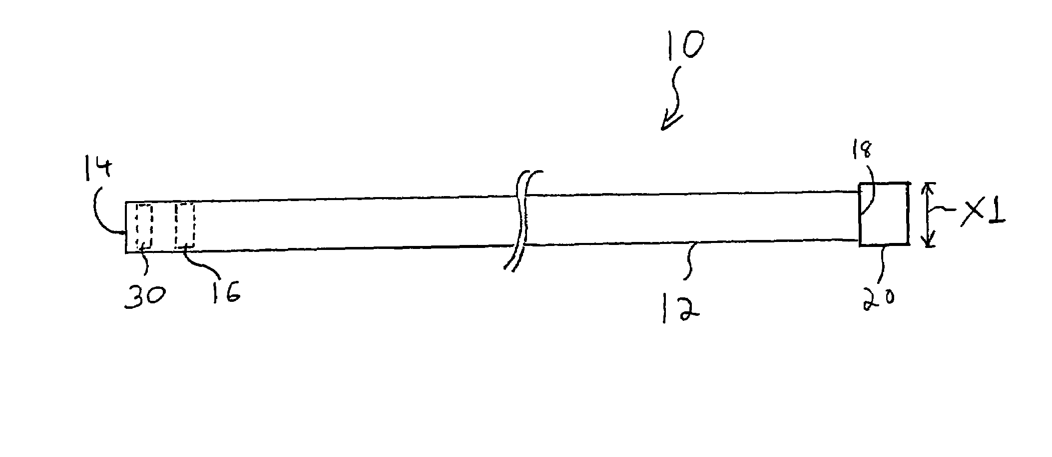
[0019]FIG. 1 is a diagram of a vision catheter system 10 formed in accordance with the present invention. The vision catheter system 10 includes a flexible catheter body 12 having a distal end 14. The vision catheter system 10 also includes a scanning mechanism and detector assembly 16, which will be described in more detail below with reference to FIG. 4.

[0020]In one embodiment, the scanning mechanism and detector assembly 16 causes a scan to occur of an image at the distal end 14 of the catheter body 12. The scanning effect causes the field of view that is sensed by the distal end 14 to effectively increase. The sensed image may be transferred to a computer or processor, and may further be recorded and / or displayed on a monitor. The vision catheter system 10 also includes a distal objective lens 30 that is placed in front of the scanning mechanism and detector assembly 16. The distal objective lens 30 is equipped with a flush port to clean the lens. In one embodiment, the distal o...


PUM


Abstract

A disposable imaging catheter that produces high resolution, color images comparable to those obtained from an endoscope. The device may also be made to function as a guidewire. The device may also include a sheath which slides over the catheter body for stiffening and which may include a working channel for accepting interventional devices, as well as LEDs to illuminate the field of view. The vision catheter system includes a detector assembly, scanning mechanism, and distal objective lens. In one embodiment, a photodetector is mated to a lens / pinhole assembly that allows the detector to read light from a small discrete point. This assembly is then scanned in raster or spiral patterns via electric wire coils that actuate a magnetic scan plate to read the area of interest. By adding a fixed objective lens, such as an aspheric lens that is attached to the distal tip of the catheter body, the field of view or acceptance angle of the system is magnified, yielding a wide angle image similar to that commonly obtained from an endoscope.

Description

FIELD OF THE INVENTION[0001]The present invention relates to medical devices, and in particular, to a catheter with imaging capabilities.BACKGROUND OF THE INVENTION[0002]Many medical interventional procedures are dependent on endoscopes to deliver diagnostic and therapeutic catheters to GI, URO, and biliary locations throughout the body. In these types of procedures, the area for maneuvering the endoscope is limited by the working channel diameter. Further limitations regarding the areas that are accessible to the endoscope are due to the physical constraints caused the size and stiffness of the endoscope. Furthermore, with regard to the comfort of the patient, endoscopic procedures are often very painful and require sedation.[0003]A typical endoscope has an illumination channel and an imaging channel, both of which may be made of a bundle of optical fibers. The illumination channel is coupled to a light source to illuminate an internal body cavity of a patient, and the imaging chan...

Claims


Application Information

Patent Timeline
Patent Type & Authority Patents(United States)
IPC IPC(8): A61B1/06A61B1/04A61B1/313
CPCA61B1/3137
Inventor BARBATO, LOUIS J.HAMM, MARK A.CHIN, YEM
Owner BOSTON SCI SCIMED INC
Why Eureka
Try Eureka