Methods and systems for detecting an analyte in a sample

a technology of analyte detection and sample, applied in the field of methods and systems for detecting analyte in a sample, can solve the problems of limited evidence of real-world performance in real-world settings, unsuitable for higher throughput applications, and 40% of patients in such settings, so as to achieve accurate measurement of infectious disease markers and dramatically reduce the interference of sample solution

Active Publication Date: 2016-12-20
BECTON DICKINSON & CO
View PDF211 Cites 3 Cited by
  • Summary
  • Abstract
  • Description
  • Claims
  • Application Information

AI Technical Summary

Benefits of technology

[0002]Point-of-care (POC) tests facilitate the diagnosis of diseases, especially in resource-limited settings where health care infrastructure is weak, and access to quality and timely medical care is a challenge. POC tests often offer rapid results, allowing for timely initiation of appropriate therapy. Importantly, POC tests can often be simple enough to be used at the primary care level and in remote settings with little to no laboratory infrastructure. POC tests thus offer certain advantages over other types of assays, such as flow cytometry-based microparticle immunoassays, which provide high accuracy and multiplexing, but are often inappropriate for POC settings due to cumbersome sample preparation and expensive instrumentation.
[0003]For example, POC tests facilitate the diagnosis or treatment of HIV / AIDS. In most resource-limited countries, eligibility for antiretroviral therapy for HIV / AIDS is based on a count of CD4+ T-lymphocytes. Traditional CD4 count technologies require that a venous blood sample be processed by a laboratory. After testing positive for HIV infection, patients either provide a blood sample immediately or are referred to another facility if the HIV counseling and testing site does not take blood samples. The blood sample is then sent to a laboratory for processing using, e.g., flow cytometry-based assays. Depending on laboratory capacity, results are typically available between 2 and 14 days after the patient has provided the sample. Patients are asked to return to the clinic to receive their results, after which they may be referred for future HIV care and / or treatment. However, roughly 40% of patients in such settings either do not provide a blood sample or do not return to the clinic to obtain their results. POC tests can facilitate the diagnosis of HIV / AIDS by simplifying the blood collection process and by making the results available while the patient is still in the clinical setting. Further, POC tests may potentially empower such patients to self-test in the privacy of their homes, especially for stigmatized diseases such as HIV / AIDS.
[0004]Most POC tests for infectious diseases offer only a single diagnosis per test. Recently, multiplexed POC tests have been developed, such as the Multiplo® POC test (MedMira Inc, NS, Canada). Multiplexed POC tests can, in principle, diagnose multiple infectious diseases, thereby offering the promise of simultaneous detection of conditions (e.g., HIV, hepatitis B and C, syphilis, etc.) with greater convenience for patients and providers. Currently, however, evidence of their performance in real world settings is limited. Rapid diagnosis of multiple infectious diseases from a single sample (e.g., a single fingerstick blood drop) using an inexpensive and facile technology available at the point-of-care would greatly improve global health outcomes.SUMMARY
[0008]Embodiments of the present disclosure enable the rapid detection of one or more analytes (e.g., 2 or more, such as 3, 4, 5, etc.) in a sample. Such detection may be qualitative, and / or quantitative. A variety of analytes may be detected by methods of the present disclosure, including analytes of biological and / or non-biological origin (e.g., chemical and / or synthetic analytes). Analytes of interest include, but are not limited to, cells, antibodies, polypeptides, polynucleotides, etc. In certain aspects, an analyte is a biomarker. Accordingly, methods of the present disclosure may facilitate the detection, monitoring, and / or diagnosis of one or more conditions in a subject from a biological sample obtained from the subject. Embodiments of the methods are amenable to POC tests to detect multiple infectious diseases from a single biological sample (e.g., a single fingerstick blood drop), using inexpensive technology.
[0017]To address the unmet needs for multiplexed point-of-care immunoassays suitable for the simultaneous detection of multiple diseases from a small volume of sample such as fingerstick whole blood, we have developed a novel approach combining multiplexed microparticle immunoassays with microfluidic assay design and inexpensive digital imaging / image processing, making an easy-to-manufacture platform for accurate measurement of infectious disease markers in whole blood with no sample preparation. The washing step normally required in conventional ELISA assays is eliminated by specifically measuring the bound fluorescence label molecules on microparticles while reducing the interference from unbound label molecules in the assay solution. Unlike conventional assays, where a combined optical signal is measured from a bulk volume of assay solution with no spatial separation, digital imaging provides detailed two-dimensional information in the sample. With imaging process and computer analysis, optical intensity from each microparticle can be measured specifically in the two-dimensional space. Another aspect of our new approach described herein is the defined height to particle size ratio. With channel height to particle ratio of 50 or less, the interference of sample and unbound fluorescence labels in the third z dimension is limited. In contract to a typical microtiter plate-based ELISA assay set up, where a combined optical signal from label molecules bound on the bottom of a microtiter plate is measured from the top of the plate, through several millimeters of assay solution containing unbound label molecules and other potentially interfering substances, our new approach involves imaging of microparticles with a thin layer of assay fluid, thus enabling the assay to be performed without the need for a washing step. As an example, in an embodiment of our invention, microparticles of 7.5 μm diameter are imaged inside a fluidic channel of 50 μm height, thus dramatically reducing the interfere with from sample solution and eliminate the need for bulk separation (washing) of the assay solution.

Problems solved by technology

Point-of-care (POC) tests facilitate the diagnosis of diseases, especially in resource-limited settings where health care infrastructure is weak, and access to quality and timely medical care is a challenge.
POC tests thus offer certain advantages over other types of assays, such as flow cytometry-based microparticle immunoassays, which provide high accuracy and multiplexing, but are often inappropriate for POC settings due to cumbersome sample preparation and expensive instrumentation.
However, roughly 40% of patients in such settings either do not provide a blood sample or do not return to the clinic to obtain their results.
Further, POC tests may potentially empower such patients to self-test in the privacy of their homes, especially for stigmatized diseases such as HIV / AIDS.
Currently, however, evidence of their performance in real world settings is limited.
However, these assays typically require a bulk liquid separation (i.e., washing) step to remove unbound label molecules before the final measurement, and are not best suited for higher throughput applications that require a high level of automation, or point-of-care applications that demand a user-friendly and low cost assay procedure.
These technologies require complicated preparation of sensing biomolecules or instrumentation and have limited applications, particularly in point-of-care applications.

Method used


Image

  • Methods and systems for detecting an analyte in a sample
  • Methods and systems for detecting an analyte in a sample
  • Methods and systems for detecting an analyte in a sample

Examples


example 1

Stably Associating Capture Beads with a Plastic Capillary Chamber

[0232]Capture beads coated with anti-mouse antibodies (BD CompBeads, BD Biosciences, San Jose, Calif.) were stably associated with a surface of a custom molded cycloolefin polymer plastic capillary chamber. To test the impact of bead properties, buffer properties, and capillary chamber surface chemistry on the ability of beads to adhere to the surface after fluid flows into the cartridge, a series of tests were performed.

[0233]The impact of the surface chemistry on the ability of the beads to adhere to a plastic surface under the given conditions was tested by attaching BD TruCount™ beads on plastic surfaces and flowing human plasma through the surface. Beads were imaged using a custom-built digital imaging system. The effect of bead properties on the ability of the capture beads to adhere to a plastic capillary chamber surface is depicted in FIG. 3, Panel B.

[0234]The effect of capillary chamber surface chemistry on th...

example 2

Detection of an Analyte in a Sample

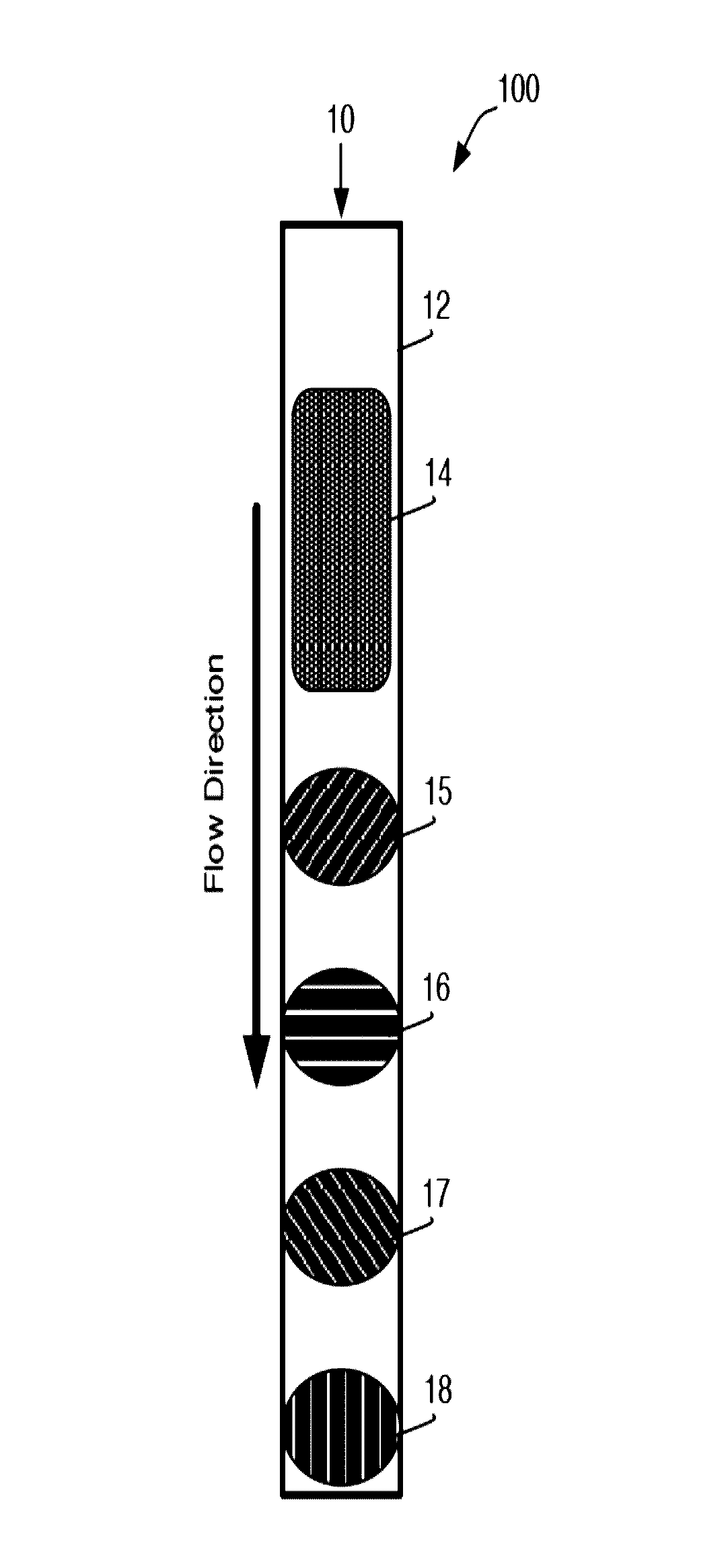
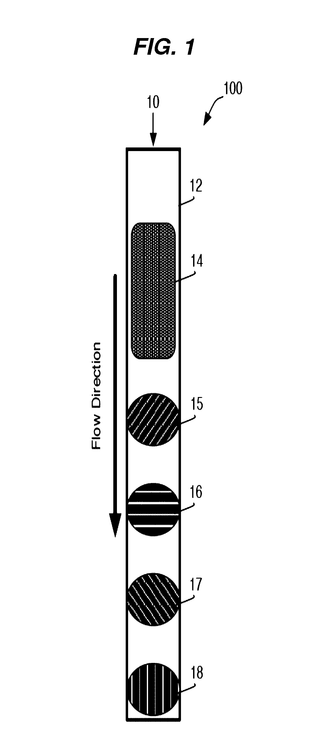
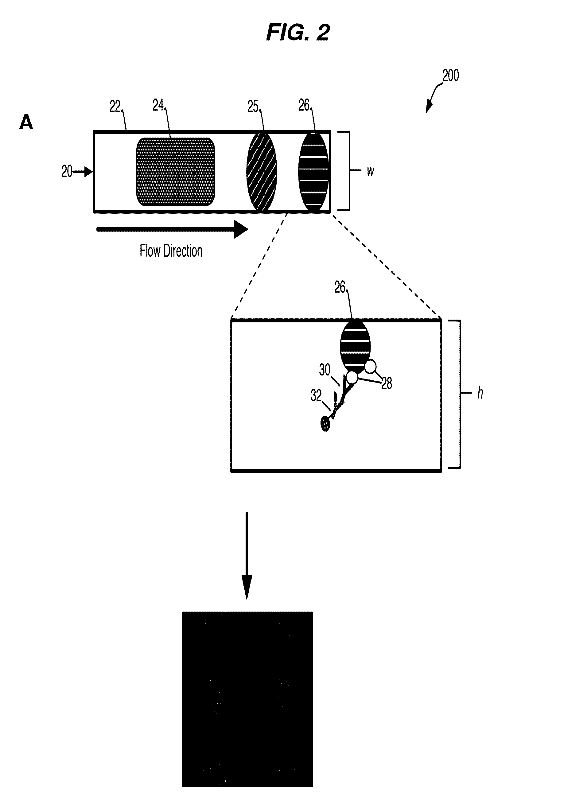
[0235]A general schematic overview of the below experiments are presented in FIG. 2, Panels A-B. Capture beads (BD CompBeads, BD Biosciences, San Jose, Calif.) were stably associated with the upper surface of a capillary chamber, as described above in Example 1. The beads were coated with anti-mouse antibodies, such that the beads would capture any mouse antibodies in the sample. The beads were labeled with mouse anti-human antibody immobilized in a spot on the upper surface of the capillary chamber. The beads stayed localized in the spot by inherent, presumably passive, interactions between the beads and the plastic chamber surface.

[0236]The capillary channel was designed such that the channel height was less than about 10 times the diameter of the capture beads, with ideal performance observed when the channel height was about 3-5 times the diameter of the capture beads.

[0237]At such dimensions, the relative low channel height reduced the backgro...


PUM

PropertyMeasurementUnit
heightaaaaaaaaaa
diameteraaaaaaaaaa
widthaaaaaaaaaa

Abstract

The present disclosure provides methods for the detection of one or more analytes in a sample. Aspects of the methods include flowing a sample (e.g., a biological sample, such as blood) through a channel comprising an analyte specific capture domain stably associated with a surface thereof, wherein the analyte specific capture domain comprises particles displaying a specific binding member for an analyte; and imaging the analyte specific capture domain to detect whether the analyte is present in the sample. Also provided are systems, devices, and kits that may be used in practicing the subject methods. Methods and compositions as described herein find use in a variety of different applications, including diagnostic applications.

Description

CROSS-REFERENCE TO RELATED APPLICATIONS[0001]Pursuant to 35 U.S.C. §119 (e), this application claims priority to the filing date of the U.S. Provisional Patent Application Ser. No. 61 / 560,752, filed Nov. 16, 2011, the disclosure of which is incorporated herein by reference.INTRODUCTION[0002]Point-of-care (POC) tests facilitate the diagnosis of diseases, especially in resource-limited settings where health care infrastructure is weak, and access to quality and timely medical care is a challenge. POC tests often offer rapid results, allowing for timely initiation of appropriate therapy. Importantly, POC tests can often be simple enough to be used at the primary care level and in remote settings with little to no laboratory infrastructure. POC tests thus offer certain advantages over other types of assays, such as flow cytometry-based microparticle immunoassays, which provide high accuracy and multiplexing, but are often inappropriate for POC settings due to cumbersome sample preparati...

Claims


Application Information

Patent Timeline
Patent Type & Authority Patents(United States)
IPC IPC(8): G01N33/543B01L3/00G01N21/64G01N35/00G01N21/03G01N21/05
CPCG01N33/54306B01L3/5027G01N21/6428G01N21/6456G01N33/54366B01L2300/0636B01L2300/087B01L2300/0809B01L2300/0838B01L2400/0406G01N21/05G01N2021/0346G01N2035/00158G01N35/00029G01N2035/00108
Inventor HUANG, WEIBORNHEIMER, SCOTT
Owner BECTON DICKINSON & CO
Why Eureka
Try Eureka