Shape memory devices and methods for reshaping heart anatomy

a memory device and heart anatomy technology, applied in the field of heart anatomy shape memory devices and methods, can solve the problems of increasing the severity of disease, the inability to reshape the heart anatomy, and the inability to fully recover from surgery, etc., to achieve the effect of increasing the cardiac output of the heart, increasing the contractibility of the ventricles, and increasing the stroke volum

Inactive Publication Date: 2007-10-23
MICARDIA CORP
View PDF47 Cites 108 Cited by
  • Summary
  • Abstract
  • Description
  • Claims
  • Application Information

AI Technical Summary

Benefits of technology

[0013]Systems, methods and devices are provided for treating heart failure patients suffering from various levels of heart dilation. Such heart dilation is treated by reshaping the heart anatomy with the use of shape memory elements. Such reshaping changes the geometry of portions of the heart, particularly the right or left ventricles, to increase contractibility of the ventricles thereby increasing the stroke volume which in turn increases the cardiac output of the heart. The shape memory elements have an original shape and at least one memory shape. The elements are implanted within the heart tissue or attached externally and / or internally to a surface of the heart when in the original shape. The elements are then activated to transition from the original shape to one of the at least one memory shapes. Transitioning of the elements cause the associated heart tissue areas to readjust position, such as to decrease the width of the ventricles. Such repositioning is maintained over time by the elements, allowing the damaging effects of heart dilation to slow in progression or reverse.
[0014]In a first aspect of the present invention, a device for reshaping the heart anatomy is provided. In preferred embodiments, the device comprises a shape memory element which is transitionable between an original shape and at least one memory shape, wherein the original shape is configured for at least partial implantation within a tissue area of the heart anatomy and the at least one memory shape is configured to apply force to the tissue area in a manner which reshapes the heart anatomy. In some embodiments, the shape memory element undergoes a deformation of at least 100% in the transition, however it may be appreciated that in some embodiments more than 100% deformation is achieved, such as several hundred percent. Further, in some embodiments, the transition substantially occurs within up to approximately 35 seconds. Such a relatively quick transition time provides advantages such as shorter procedure time and ease of use.
[0019]In a second aspect of the present invention, a method of reshaping heart anatomy is provided. In preferred embodiments, the method includes providing a shape memory element which is transitionable between an original shape and at least one memory shape, implanting the shape memory element having its original shape at least partially within a tissue area of the heart anatomy, and activating the shape memory element causing transition from its original shape to one of the at least one memory shapes while the element is at least partially implanted, wherein the transition causes the tissue area to move in a manner which reshapes the heart anatomy. Typically, the tissue area is located within a wall of a ventricle and reshaping the heart anatomy comprises reducing a width of the ventricle. And, activating may comprise providing a change in temperature, application of light, change in ionic concentration, a change in pH, application of an electric field, application of a magnetic field, application of ultrasound or a combination of these.

Problems solved by technology

In such cases, the heart may enlarge to such an extent that the adverse consequences of heart enlargement continue after the viral infection has passed and the disease continues its progressively debilitating course.
The thinned portion of the myocardium often is functionally impaired, and other portions of the myocardium attempt to compensate.
Cardiac remodeling often subjects the heart wall to increased wall tension or stress, which further impairs the heart's functional performance.
A vicious cycle arises, leading to increasing distension of the heart and consequent heart insufficiency.
Unfortunately, not enough hearts are available for transplant to meet the needs of heart failure patients.
While the availability of donor hearts has historically increased, the rate of increase is slowing dramatically.
Even if the risks and expense of heart transplant could be tolerated, this treatment option is becoming increasingly unavailable.
Further, many patients do not qualify for heart transplant for failure to meet any one of a number of qualifying criteria.
This is a radical and experimental procedure subject to substantial controversy.
Furthermore, the procedure is highly invasive, risky and expensive and commonly includes other expensive procedures (such as a concurrent heart valve replacement).
Even though cardiomyoplasty has demonstrated symptomatic improvement, studies suggest the procedure only minimally improves cardiac performance.
In addition, the procedure is highly invasive requiring harvesting a patient's muscle and an open chest approach (i.e., sternotomy) to access the heart.
Furthermore, the procedure is expensive, especially for those using a paced muscle which require costly pacemakers.
The cardiomyoplasty procedure is also complicated.
For example, it is difficult to adequately wrap the muscle around the heart with a satisfactory fit.
Also, if adequate blood flow is not maintained to the wrapped muscle, the muscle may necrosis.
Finally, the muscle may fibrose and adhere to the heart causing undesirable constraint on the contraction of the heart during systole.
However, these designs require invasive open chest surgery, significant handling of the heart, and have not seen widespread success.

Method used


Image

  • Shape memory devices and methods for reshaping heart anatomy
  • Shape memory devices and methods for reshaping heart anatomy
  • Shape memory devices and methods for reshaping heart anatomy

Examples


Embodiment Construction

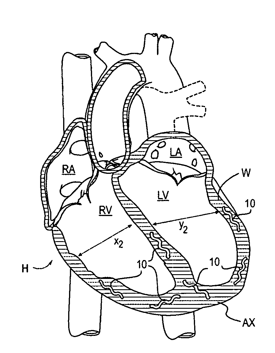
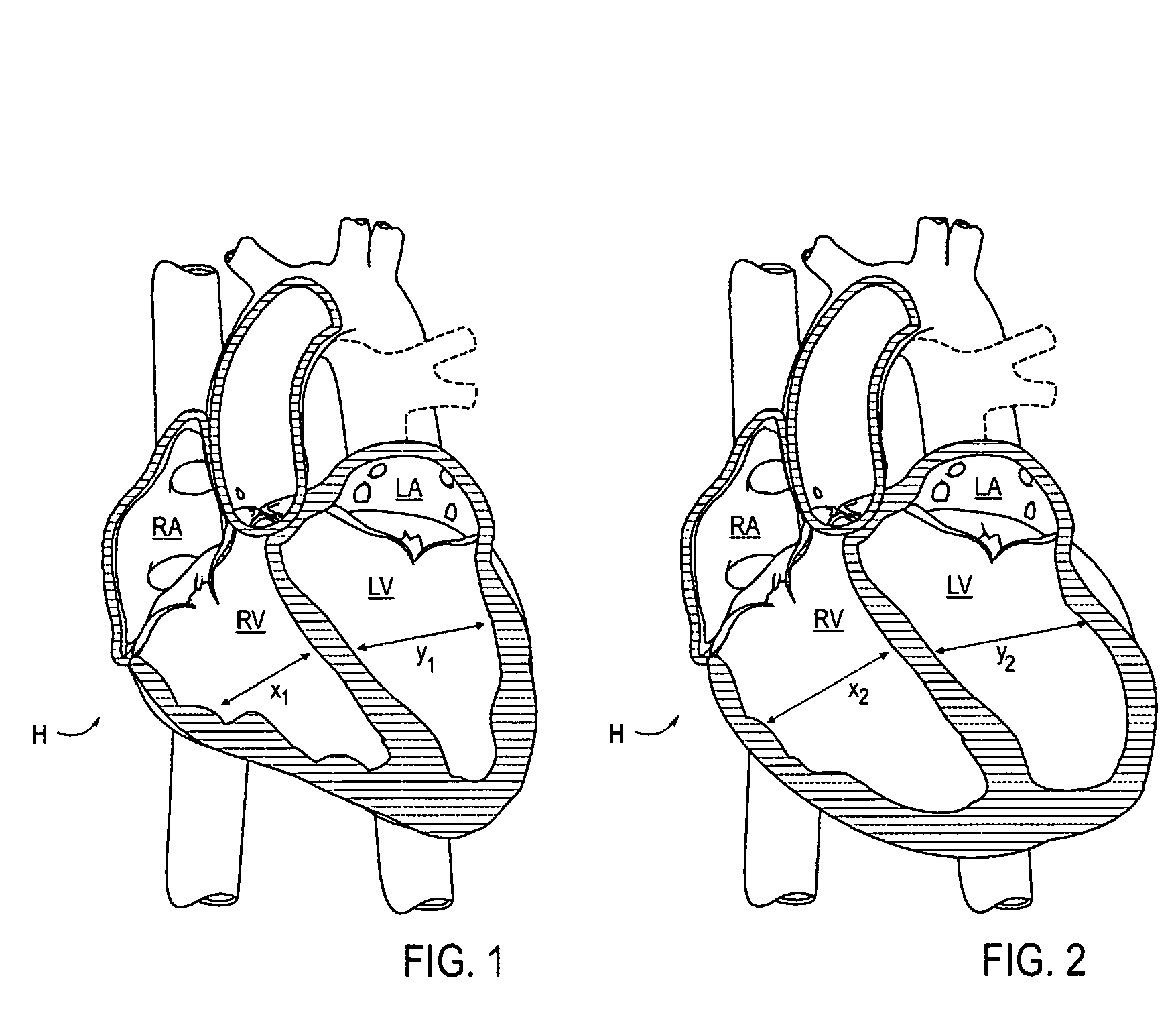
[0040]FIG. 1 provides a cross-sectional illustration of a heart H of a normal patient. The cross-sectional view shows the right atrium RA, right ventricle RV, left atrium LA and left ventricle LV. The right ventricle RV and left ventricle LV have a width of x1 and y1 respectively. FIG. 2 provides a cross-sectional illustration of a heart H of a patient with heart disease wherein the geometry of the ventricles RV, LV have dilated. As shown, the right ventricle RV and left ventricle LV have increased widths of x2 and y2 respectively. The increased widths x2, y2 result in poor cardiac output from the left ventricle LV and / or the right ventricle RV. Cardiac output (CO) is defined as:

CO=HR×SV

whereas

HR=heart rate (beats per minute)

SV=stroke volume (liters per beat)

Ejection Fraction (EF) is the fraction of blood ejected by a ventricle relative to its end-diastolic volume. Therefore, EF is calculated from:

EF=(SV / EDV)*100

whereas

EDV=end-diastolic volume

Ejection fraction is most commonly meas...


PUM


Abstract

Systems, methods and devices are provided for treating heart failure patients suffering from various levels of heart dilation. Such heart dilation is treated by reshaping the heart anatomy with the use of shape memory elements. Such reshaping changes the geometry of portions of the heart, particularly the right or left ventricles, to increase contractibility of the ventricles thereby increasing the stroke volume which in turn increases the cardiac output of the heart. The shape memory elements have an original shape and at least one memory shape. The elements are implanted within the heart tissue or attached externally and / or internally to a surface of the heart when in the original shape. The elements are then activated to transition from the original shape to one of the at least one memory shapes. Transitioning of the elements cause the associated heart tissue areas to readjust position, such as to decrease the width of the ventricles. Such repositioning is maintained over time by the elements, allowing the damaging effects of heart dilation to slow in progression or reverse.

Description

CROSS-REFERENCES TO RELATED APPLICATIONS[0001]This application claims the priority of U.S. Provisional Patent Application No. 60 / 588,254, filed on Jul. 15, 2004, incorporated herein by reference for all purposes.[0002]This application is also related to U.S. patent application Ser. No. 11 / 142,127, filed on the same day as the instant application and incorporated herein by reference for all purposes.STATEMENT AS TO RIGHTS TO INVENTIONS MADE UNDER FEDERALLY SPONSORED RESEARCH OR DEVELOPMENT[0003]Not ApplicableREFERENCE TO A “SEQUENCE LISTING,” A TABLE, OR A COMPUTER PROGRAM LISTING APPENDIX SUBMITTED ON A COMPACT DISK[0004]Not ApplicaqbleBACKGROUND OF THE INVENTION[0005]Heart failure is a common course for the progression of many forms of heart disease. Heart failure may be considered to be the condition in which an abnormality of cardiac function is responsible for the inability of the heart to pump blood at a rate commensurate with the requirements of the metabolizing tissues, or ca...

Claims


Application Information

Patent Timeline
Patent Type & Authority Patents(United States)
IPC IPC(8): A61F2/00
CPCA61B17/00234A61B17/06166A61B17/0644A61B2017/06052A61B2017/00243A61B2017/00867A61B17/068
Inventor MOADDEB, SHAHRAMSHAOLIAN, SAMUEL M.SHAOULIAN, EMANUELRHEE, RICHARDANDERSON, STEVEN C.
Owner MICARDIA CORP
Why Eureka
Try Eureka