Eureka AIR delivers breakthrough ideas for toughest innovation challenges, trusted by R&D personnel around the world.

Computer aided diagnosis of an image set

Active Publication Date: 2007-11-13
GE MEDICAL SYST GLOBAL TECH CO LLC
View PDF71 Cites 55 Cited by
  • Summary
  • Abstract
  • Description
  • Claims
  • Application Information

AI Technical Summary

Benefits of technology

[0011]The above discussed and other drawbacks and deficiencies are overcome or alleviated by a method for computer aided processing of dual or multiple energy images including employing a data source, the data source including a dual or multiple energy image set including a high energy image, a low energy image, a bone image, and a soft tissue image, defining a region of interest within an image from the dual or multiple energy image set, extracting feature measures from the region of interest, and reporting at least one of the feature measures on the region of interest.

Problems solved by technology

In addition, due to the underlying and overlying structures relevant to the pathologies of interest, visual assessment can be difficult.
Subtle rib fractures, calcifications, and metastatic bone lesions (metastases) in the chest can be difficult to detect on a standard chest X-ray.

Method used


Image

  • Computer aided diagnosis of an image set
  • Computer aided diagnosis of an image set
  • Computer aided diagnosis of an image set

Examples


Embodiment Construction

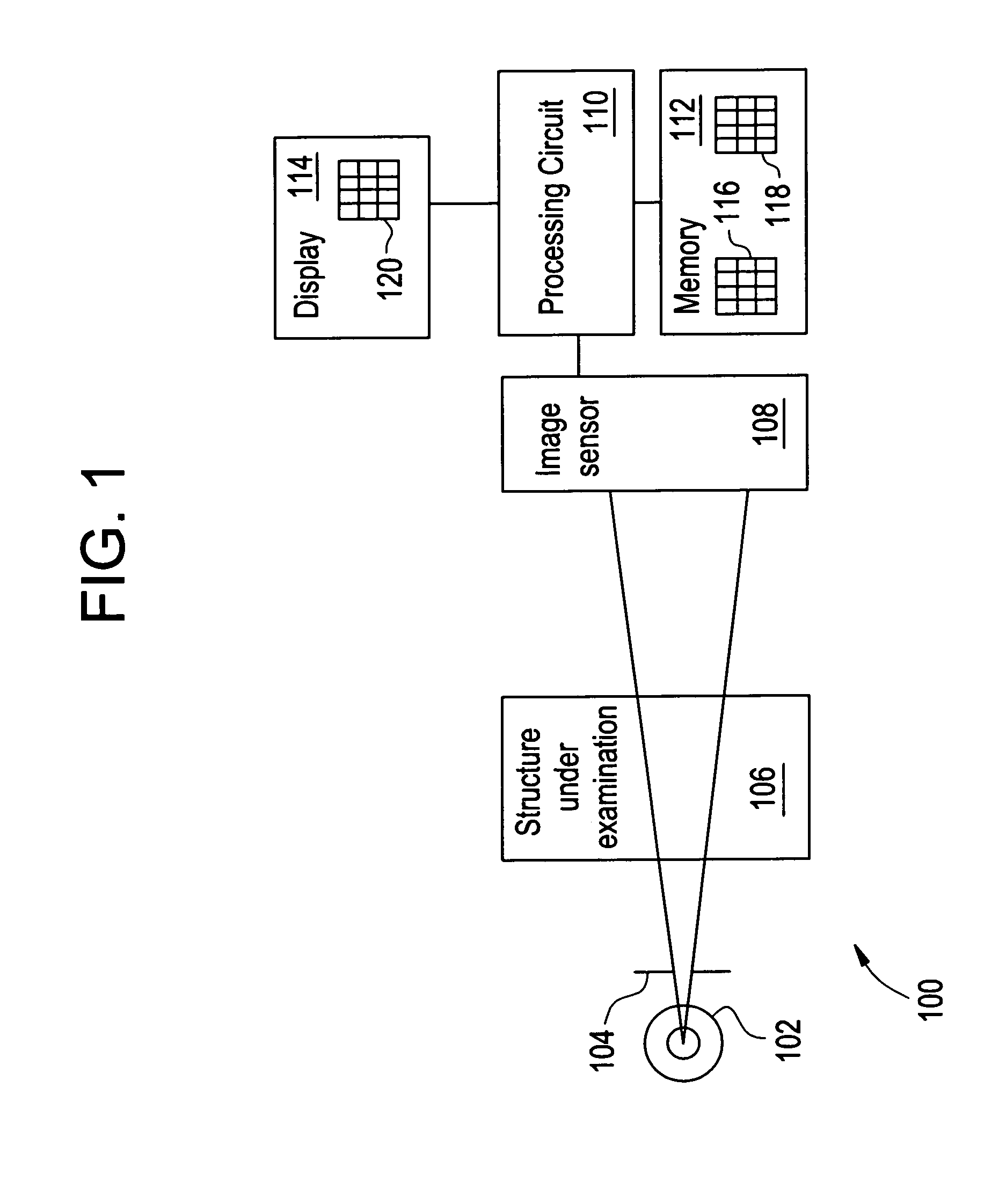
[0040]FIG. 1 illustrates an exemplary X-ray imaging system 100. The imaging system 100 includes an X-ray source 102 and a collimator 104, which subject structure under examination 106 to X-ray photons. As examples, the X-ray source 102 may be an X-ray tube, and the structure under examination 106 may be a human patient, test phantom or other inanimate object under test. The X-ray source 102 is able to generate photons at a first energy level and at least a second energy level different than the first energy level. Multiple, more than two, energy levels are also within the scope of this method and system.

[0041]The X-ray imaging system 100 also includes an image sensor 108 coupled to a processing circuit 110. The processing circuit 110 (e.g., a microcontroller, microprocessor, custom ASIC, or the like) is coupled to a memory 112 and a display 114. The display 114 may include a display device, such as a touch screen monitor with a touch-screen interface. As is known in the art, the sys...


PUM


Abstract

A method, system, and storage medium for computer aided processing of an image set includes employing a data source, the data source including an image set acquired from X-ray projection imaging, x-ray computed tomography, or x-ray tomosynthesis, defining a region of interest within one or more images from the image set, extracting feature measures from the region of interest, and reporting at least one of the feature measures on the region of interest. The method may be employed for identifying bone fractures, disease, obstruction, or any other medical condition.

Description

CROSS REFERENCE TO RELATED APPLICATIONS[0001]This application is a continuation-in-part of U.S. patent application Ser. No. 10 / 063,819, filed on May 15, 2002, pending, which is hereby incorporated by reference in its entirety.BACKGROUND OF THE INVENTION[0002]This invention generally relates to computer aided detection and diagnosis (CAD) of an image set. More particularly, this invention relates to a method and system for computer aided detection and diagnosis of dual energy (“DE”) or multiple energy images, as well as of radiographic images, computed tomography images, and tomosynthesis images.[0003]The classic radiograph or “X-ray” image is obtained by situating the object to be imaged between an X-ray emitter and an X-ray detector made of photographic film.[0004]Emitted X-rays pass through the object to expose the film, and the degree of exposure at the various points on the film are largely determined by the density of the object along the path of the X-rays.[0005]It is now comm...

Claims


Application Information

Patent Timeline
IPC IPC(8): G06K9/00A61B6/00A61B5/00A61B6/03G06F7/40G06F17/50G06K9/34G06T1/00G06T3/00G06T5/30G06T7/00G06T11/00G16H30/20G16H50/20
CPCA61B6/482G06F19/321G06T11/005G06T7/0012G06T2211/408G06T2207/30008A61B6/032A61B6/502A61B6/505G16H50/20G16H30/20A61B6/03
Inventor UPPALURI, RENUKARADER, AMBER ELAINEAVINASH, GOPAL B.THOMAS, CARSON HALESABOL, JOHN MICHAELJABRI, KADRI NIZAR
Owner GE MEDICAL SYST GLOBAL TECH CO LLC
Why Eureka
Eureka Blog
Learn More