Method and apparatus for the three-dimensional presentation of an examination region of a patient in the form of a 3D reconstruction image

a three-dimensional reconstruction and patient technology, applied in the field of three-dimensional reconstruction of the examination region of a patient, can solve the problems of difficult diagnosis, examination or treatment, and difficult anatomical conditions, and achieve the effect of improving the quality of li

Active Publication Date: 2007-11-27
SIEMENS HEALTHCARE GMBH
View PDF24 Cites 107 Cited by
  • Summary
  • Abstract
  • Description
  • Claims
  • Application Information

AI Technical Summary

Benefits of technology

[0006]An object of the present invention is to provide a method and apparatus for the three-dimensional presentation of an examination region of a patient in the form of a 3D reconstruction image, wherein the aforementioned disadvantages are alleviated.

Problems solved by technology

Particularly when the 3D reconstruction image is employed in the context of ongoing intervention, difficulties can occur due to anatomical conditions that have changed since the preoperative exposure, i.e. the 3D reconstruction image that is reconstructed based on the preoperative image data no longer represents the current anatomical or positional conditions.
Ambiguities arise therefrom for the attending physician that can make the diagnosis, examination or treatment more difficult, particularly the intervention treatment.

Method used


Image

  • Method and apparatus for the three-dimensional presentation of an examination region of a patient in the form of a 3D reconstruction image
  • Method and apparatus for the three-dimensional presentation of an examination region of a patient in the form of a 3D reconstruction image
  • Method and apparatus for the three-dimensional presentation of an examination region of a patient in the form of a 3D reconstruction image

Examples


Embodiment Construction

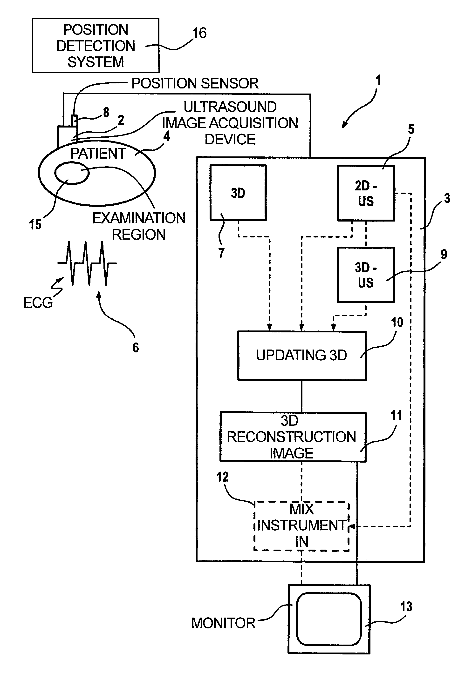
[0022]In a schematic illustration, FIG. 1 shows an inventive examination and / or treatment device 1 having an ultrasound image acquisition device 2 as well as a control and processing device 3 that controls the operation of the ultrasound image acquisition device 2 and also undertakes the processing, editing and analysis of the image data. A set of 2D ultrasound images of an examination region—the heart of a patient 4 in this case—that are forwarded to the control and processing device 3, are acquired with the ultrasound device 2. In the illustrated example, the acquisition of the image data representing 2D ultrasound images ensues with triggering by an ECG 6 that is recorded in parallel, since the examination region 15 is a rhythmically moving organ, namely the heart. The ECG data are likewise forwarded to the control and processing device 3.

[0023]A position sensor 8 with which the spatial position of the ultrasound acquisition device 2, and thus the respective spatial position of e...


PUM


Abstract

In a method and apparatus for the three-dimensional presentation of an examination region of a patient in the form of a 3D reconstruction image, a preoperatively acquired 3D image dataset of the examination region is employed in a medical procedure, datasets representing a number of 2D ultrasound images of the examination region are acquired, the preoperative 3D image dataset is updated using the datasets representing 2D ultrasound images, and the 3D reconstruction image is generated on the basis of the updated 3D image dataset.

Description

BACKGROUND OF THE INVENTION[0001]1. Field of the Invention[0002]The present invention is directed to a method and apparatus for the three-dimensional presentation of an examination region of a patient in the form of a 3D reconstruction image.[0003]2. Description of the Prior Art[0004]The three-dimensional presentation of an examination region of a patient in the form of a 3D reconstruction image is becoming increasingly important in the framework of medical examinations or treatments. Examples are minimally invasive treatments using endoscopes, laparoscopes or catheters that are respectively introduced into the examination region of the patient via a small body opening. On the basis of the 3D reconstruction image, the physician is provided with a three-dimensional view of the examination region, for example of the heart, which is useful to the physician for the navigation of the medical instrument. Such 3D reconstruction images, however, also are useful in instances wherein no instr...

Claims


Application Information

Patent Timeline
Patent Type & Authority Patents(United States)
IPC IPC(8): A61B5/05A61B8/13A61B19/00
CPCA61B8/13A61B8/543A61B2019/5289A61B2090/364
Inventor CAMUS, ESTELLEDITT, HENDRIKKILLMANN, REINMARRAHN, NORBERTWACH, SIEGFRIED
Owner SIEMENS HEALTHCARE GMBH
Why Eureka
Try Eureka