Feature quantification from multidimensional image data

a multi-dimensional image and feature technology, applied in image enhancement, instruments, applications, etc., can solve the problems of reducing the perfusion capacity of the affected organ, reducing the capacity of the vessel to convey blood, and continuing serious limitations in the available approaches for assessment of vascular disease, so as to facilitate the assessment of stenosis

Inactive Publication Date: 2008-02-19
GENERAL ELECTRIC CO
View PDF3 Cites 141 Cited by
  • Summary
  • Abstract
  • Description
  • Claims
  • Application Information

AI Technical Summary

Benefits of technology

[0034]As an aid for practicing the present invention, also disclosed herein are a method, an apparatus, and a computer-readable medium storing a program in which volumetric images are reformatted into one or more two-dimensional (2D) representations at selected plane orientations. The selected orientations of the reformatted 2D image planes can be chosen to provide desirable orientations of a feature or features depicted in the reformatted images. In a desirable embodiment, the invention provides an approach to reformatting volumetric images of blood vessels onto special 2D grids which facilitate assessment of stenosis.

Problems solved by technology

For example, serious limitations have continued to exist in the available approaches for assessment of vascular disease.
Stenosis reduces the vessel's capacity to convey blood and thus can seriously reduce perfusion in the affected organ.
Catheterization procedures are effective but are also highly invasive.
However, as with conventional angiographic catheterization, IVUS techniques are highly invasive.
These factors, and the need for a skilled operator to collect reliable data, raise important issues for reliable and repeatable assessment of vascular disease.
An image transverse to the vessel's longitudinal axis would be unsuitable for this purpose, because diameters along the longitudinal axis could not be compared.
However, the foregoing observations illustrate some of the sources of assessment error that are associated with estimating the degree of stenosis based on lumen diameters.
However, frequently the reconstructed images will depict the structure of interest in an inconvenient orientation for purposes of examining particular structural features.
The general result has been reduced precision for the assessment to be performed.
The desired techniques would be readily applied in a range of areas where quantification of structural dimensions or other extensional features is needed and is currently unfeasible or subject to poorly constrained sources of error.
Further, because any necessary contrast medium could be injected into the peripheral vasculature, the desired technique would be only minimally invasive.

Method used


Image

  • Feature quantification from multidimensional image data
  • Feature quantification from multidimensional image data
  • Feature quantification from multidimensional image data

Examples


Embodiment Construction

[0051]Reference will now be made in detail to the presently preferred embodiments of the invention, examples of which are illustrated in the accompanying drawings, wherein like reference numerals refer to like elements throughout.

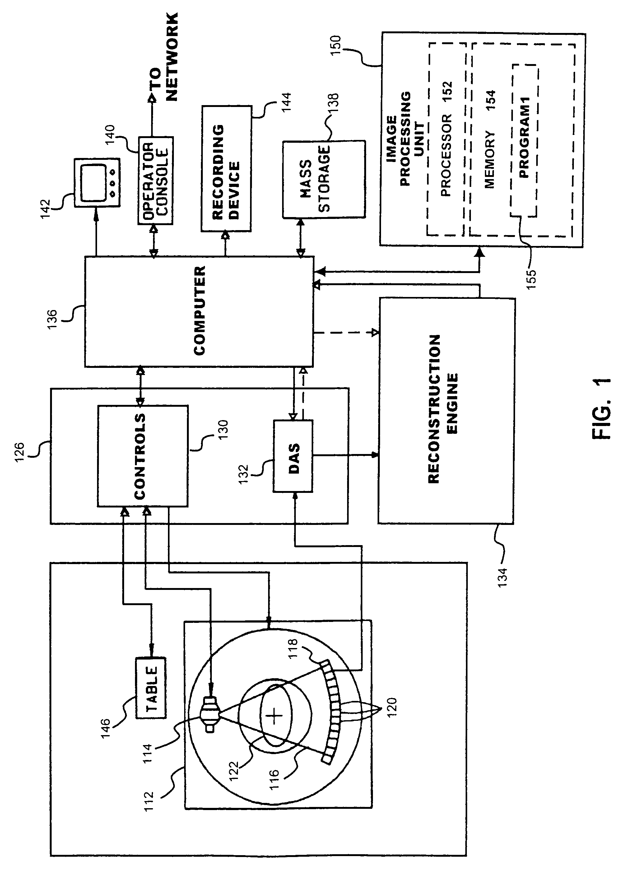
[0052]FIG. 1 is a schematic illustration showing the major components of a CT imaging system 100 in which the present invention may be incorporated. The CT system 100 desirably comprises a source-detector assembly 110. In some systems, such as so-called third generation scanners (“fan beam” systems) and some helical scanners, the assembly 110 may comprise a gantry 112. An X-ray source 114, such as a typical X-ray tube, produces a beam 116 of illuminating x-rays from which projection data may be obtained.

[0053]For purposes of clarity, the following description will specifically describe the fan beam case. Those of ordinary skill in the art will readily recognize and appreciate the occasional differences between third generation and fourth generation systems ...


PUM


Abstract

Techniques, hardware, and software are provided for quantification of extensional features of structures of an imaged subject from image data representing a two-dimensional or three-dimensional image. In one embodiment, stenosis in a blood vessel may be quantified from volumetric image data of the blood vessel. A profile from a selected family of profiles is fit to selected image data. An estimate of cross sectional area of the blood vessel is generated based on the fit profile. Area values may be generated along a longitudinal axis of the vessel, and a one-dimensional profile fit to the generated area values. An objective quantification of stenosis in the vessel may be obtained from the area profile. In some cases, volumetric image data representing the imaged structure may be reformatted to facilitate the quantification, when the structural feature varies along a curvilinear axis. A mask is generated for the structural feature to be quantified based on the volumetric image data. A curve representing the curvilinear axis is determined from the mask by center-finding computations, such as moment calculations, and curve fitting. Image data are generated for oblique cuts at corresponding selected orientations with respect to the curvilinear axis, based on the curve and the volumetric image data. The oblique cuts may be used for suitable further processing, such as image display or quantification.

Description

CROSS REFERENCE TO RELATED APPLICATIONS[0001]This application a continuation of application Ser. No. 09 / 648,958, filed Aug. 25, 2000, now abandoned which parent application is related to Provisional Application U.S. Ser. No. 60 / 197,208, filed Apr. 14, 2000 and Provisional Application U.S. Ser. No. 60 / 166,499, filed Nov. 19, 1999, in the U.S. Patent and Trademark Office. The contents of the aforementioned Provisional Application are incorporated herein by reference, and the benefit of priority to the same Provisional Application is claimed under 35 U.S.C. §119(e).BACKGROUND OF THE INVENTION[0002]The present invention relates to quantitative analysis of image data. More specifically, the invention relates to quantification of extensional features of structures of an imaged subject from image data representing a two-dimensional or three-dimensional image. In a particular embodiment, the invention may be used for analysis of tomographic image data for quantification of stenosis in vascu...

Claims


Application Information

Patent Timeline
Patent Type & Authority Patents(United States)
IPC IPC(8): G06K9/00G06K9/46A61B6/00G06T11/00
CPCG06T7/0012A61B6/508G06T7/602A61B6/541G06T2207/10081G06T2207/30101G06T7/62
Inventor EDIC, PETER MICHAELYAVUZ, MEHMETISHAQUE, AHMAD NADEEM
Owner GENERAL ELECTRIC CO
Why Eureka
Try Eureka