
Method of magnetic resonance investigation of a sample using a nuclear spin polarised MR imaging agent

a nuclear spin polarised mr imaging and magnetic resonance imaging technology, applied in the field of magnetic resonance imaging (mri), can solve the problems of comparatively short tsub>1 /sub>values of nuclei and less than optimal image quality

Inactive Publication Date: 2008-03-18
OXFORD INSTR MOLECULAR BIOTOOLS
View PDF10 Cites 0 Cited by
  • Summary
  • Abstract
  • Description
  • Claims
  • Application Information

AI Technical Summary

Problems solved by technology

Since superconducting magnets and complex magnet construction are required for large cavity high field strength magnets, these are expensive.
However there are no particular advantages to using the very high field strengths where noise from the patient begins to dominate over electronic noise (generally at field strengths where the resonance frequency of the imaging nucleus is 1 to 20 MHz) and accordingly the use of hyperpolarised “contrast agents opens the possibility of high performance imaging using low cost, low field strength magnets.
one problem with these previously described techniques is that whilst the value of the gyromagnetic ratio, γ, for hydrogen is 42.6 MHz / T, it is much lower for both carbon and nitrogen, at 10.7 MHz / T and 4.3 MHz / T, respectively.
A further drawback in using 13C or 15N-based contrast medium, particularly in angiography, relates to the gradient power that is required for the MRI.
Thus, if hyperpolarised contrast media containing non-proton imaging nuclei, particularly 13C or 15N nuclei, are to be used in combination with fast imaging sequences, there will be a less than optimal image quality, due to the lower values of the gyromagnetic ratio of the non-proton imaging nuclei.
Furthermore, a problem in using nuclei with relatively high gyromagnetic ratios in ex vivo polarisation techniques is that such nuclei have comparatively short T1 values.

Method used


Image

  • Method of magnetic resonance investigation of a sample using a nuclear spin polarised MR imaging agent
  • Method of magnetic resonance investigation of a sample using a nuclear spin polarised MR imaging agent
  • Method of magnetic resonance investigation of a sample using a nuclear spin polarised MR imaging agent

Examples


Embodiment Construction

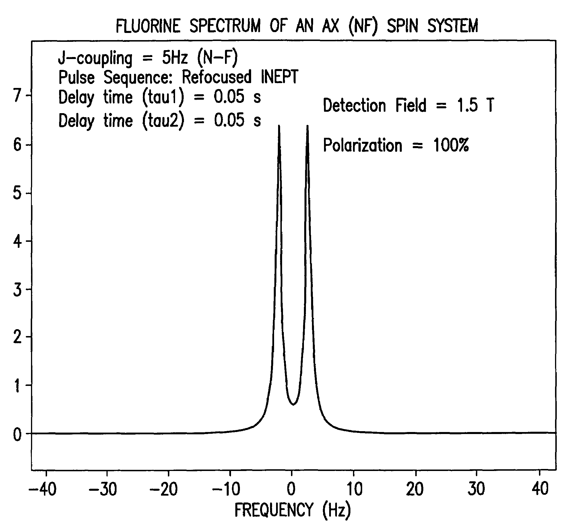
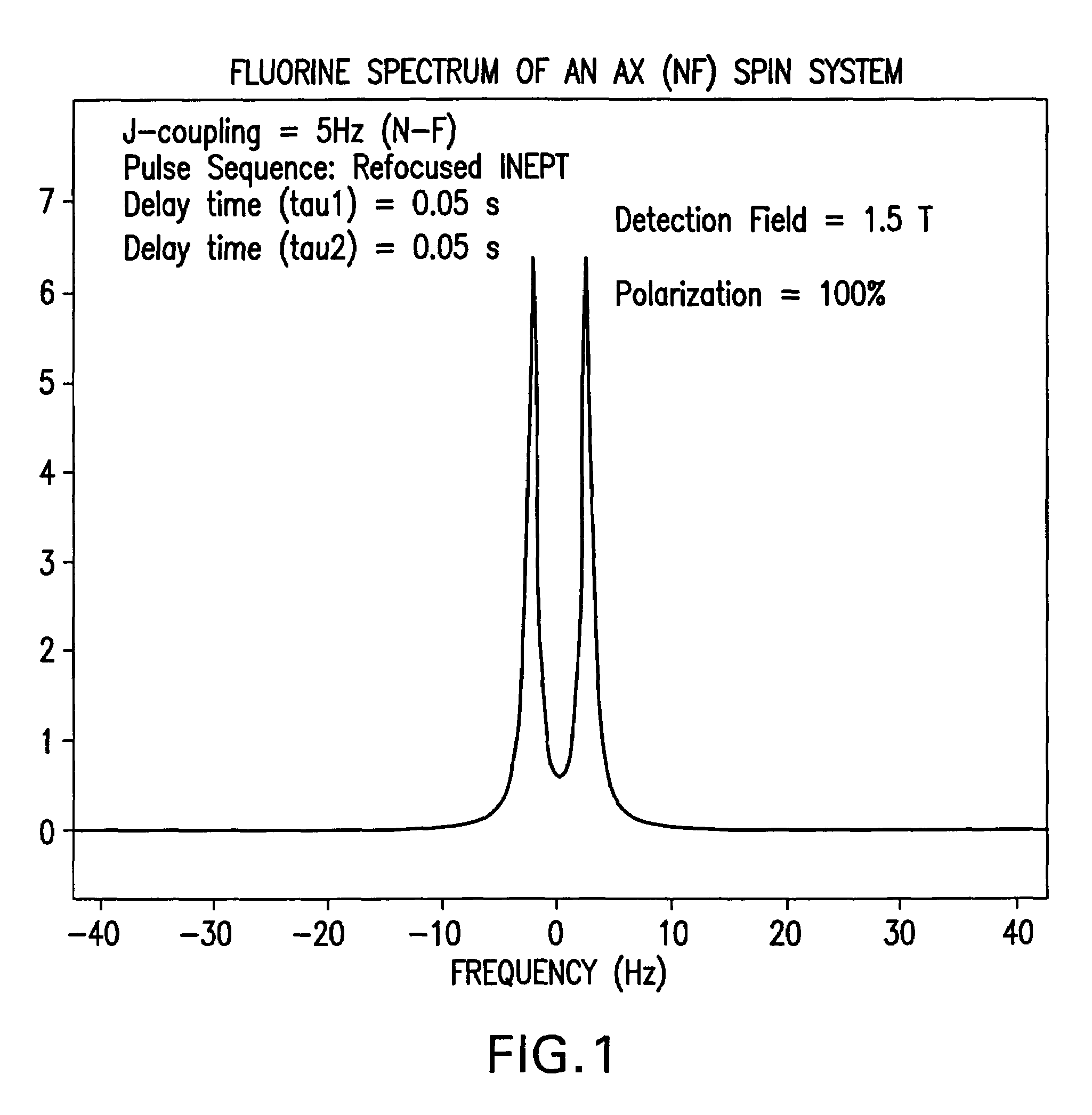
[0024]The present invention thus relates in one aspect to a method whereby the above-mentioned drawbacks are addressed by using a technique in which after production of a contrast media containing hyperpolarised nuclei, preferably 13C or 15N nuclei, the media is injected into the patient, and the patient is then subjected to a pulse sequence which transfers polarisation from the hyperpolarised nuclei, e.g. the 13C or 15N nuclei, to nuclei having a higher value of the gyromagnetic ratio, e.g. 1H, 19F or 31P nuclei, which then serve as the imaging nuclei for image generation.

[0025]Thus viewed from one aspect the present invention provides a method of magnetic resonance investigation of a sample, preferably a human or non-human animal body (e.g. a mammalian, reptilian or avian body), said method comprising:[0026]i) obtaining a MR imaging agent containing in its molecular structure at least one storage non-zero nuclear spin nuclei;[0027]ii) nuclear spin polarising said storage nuclei in...


PUM


Abstract

The present invention provides a method of contrast enhanced magnetic resonance imaging of a sample, said method comprising: a) administering a hyperpolarised MR contrast agent comprising non-zero nuclear spin nuclei into said sample for fluid dynamic investigations of the vasculature, b) exposing said sample or part of the sample to radiation of a frequency selected to excite nuclear spin transitions in said non-zero nuclear spin nuclei, c) detecting MR signals from said sample using any suitable manipulation method including pulse sequences. The invention also provides novel compounds.

Description

[0001]This application claims priority from Continuation of PCT / GB01 / 04096 filed on Sep. 11, 2001, which claims priority from Provisional Application 60 / 259,933 filed on Jan. 5, 2001 and to United Kingdom Application No. 0022341.2 filed on Sep. 12, 2000 all of which are hereby incorporated by reference.FIELD OF INVENTION[0002]The present invention relates to methods of magnetic resonance imaging (MRI), in particular to a technique involving polarisation transfer between different nuclei with different gyromagnetic ratios (V).BACKGROUND OF THE INVENTION[0003]Magnetic resonance imaging is a diagnostic technique that has become particularly attractive to physicians as it is non-invasive and does not involve exposing the patient under study to potentially harmful radiation such as X-rays.[0004]In order to achieve effective contrast between MR images of different tissue types, it has long been known to administer to the subject MR contrast agents (e.g. paramagnetic metal species) which a...

Claims


Application Information

Patent Timeline
IPC IPC(8): A61B5/05A61B5/055G01R33/28A61K49/00A61K49/10G01R33/483
CPCA61K49/08G01R33/5601G01R33/28
Inventor PETERSSON, STEFANAXELSSON, OSKARJOHANNESON, HAUKUR
Owner OXFORD INSTR MOLECULAR BIOTOOLS
Learn More