

Charged particle beam equipment and charged particle microscopy
a charge beam and equipment technology, applied in the direction of material analysis using wave/particle radiation, instruments, nuclear engineering, etc., can solve the problems of specimens that cannot be used for high magnification calibration, cannot be applied to the entire range of observation magnification of electron microscopes, and cannot be used for m calibration, etc., to reduce the effect of specimen dri
- Summary
- Abstract
- Description
- Claims
- Application Information
AI Technical Summary
Benefits of technology
Problems solved by technology
Method used
Image

Examples
Embodiment Construction
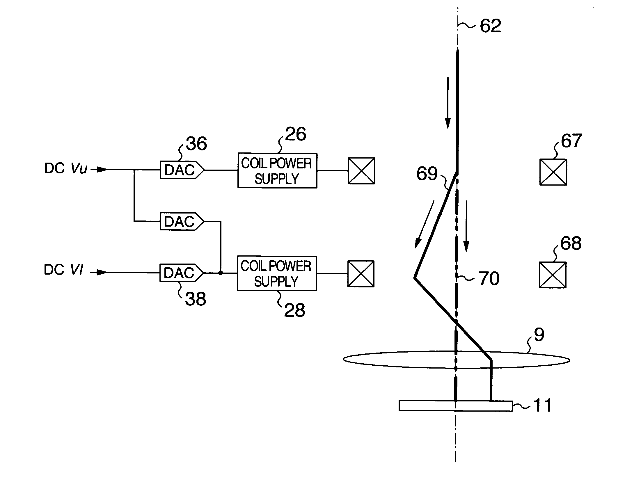
[0035]Description will be made of an embodiment of the present invention with reference to the accompanying drawings. The present invention is applicable to cases where the charged particle beam is an ion beam or an electron beam; however, the present invention will be described referring to a case where the charged particle beam is an electron beam in the following. In addition, the lens system, the beam deflector and the scanner may be either of the type using an electric field or of the type using a magnetic field; however, description will be made of a case where those devices of the magnetic field type are used. It does not matter at all whether there is a projection lens under a specimen or how many stage the projection lens may be.
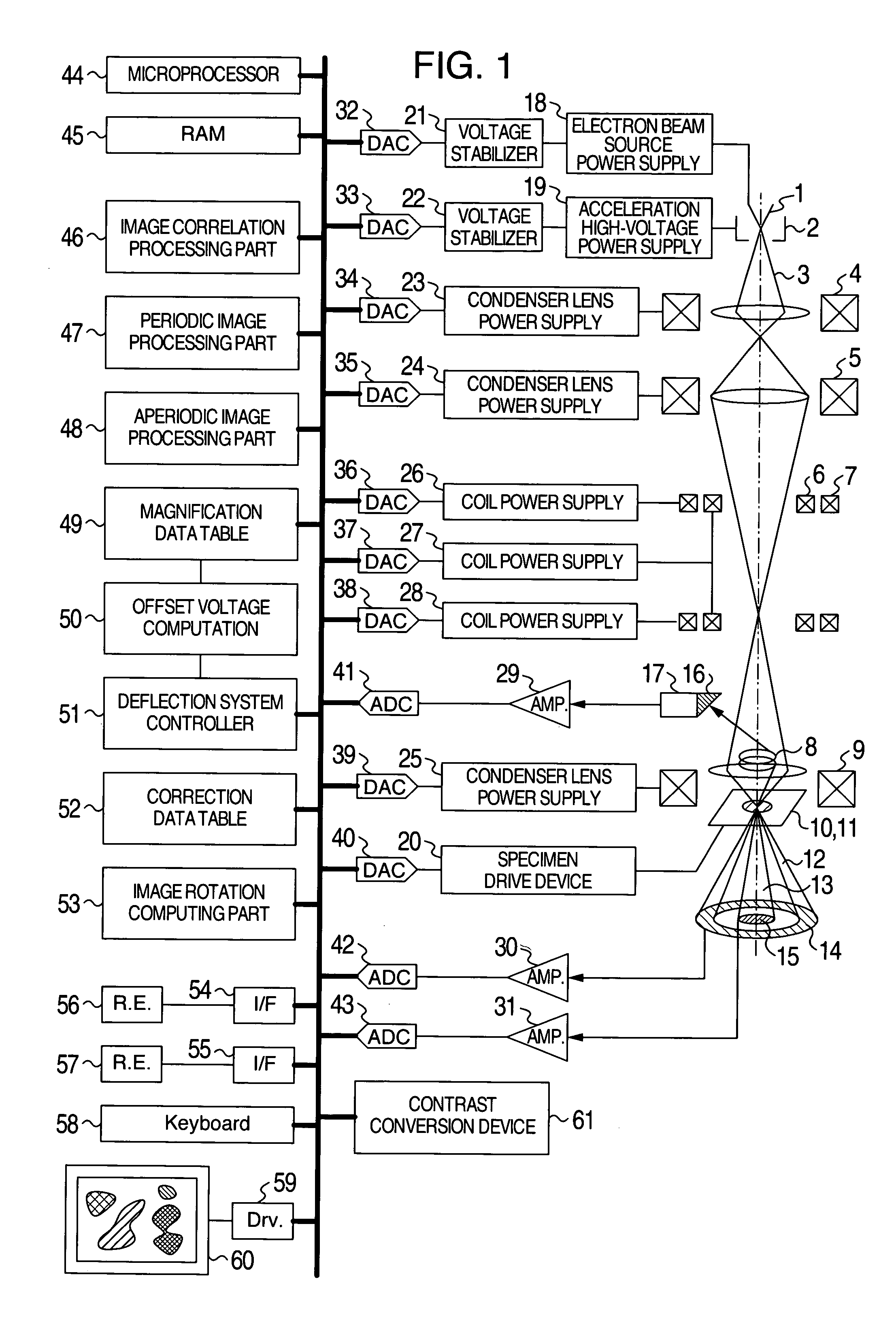
[0036]FIG. 1 is a schematic function block diagram as an example of charged particle beam equipment according to the present invention.
[0037]An electron beam (charged particle beam) 3 emitted from an electron beam source (charged particle beam sourc...
PUM
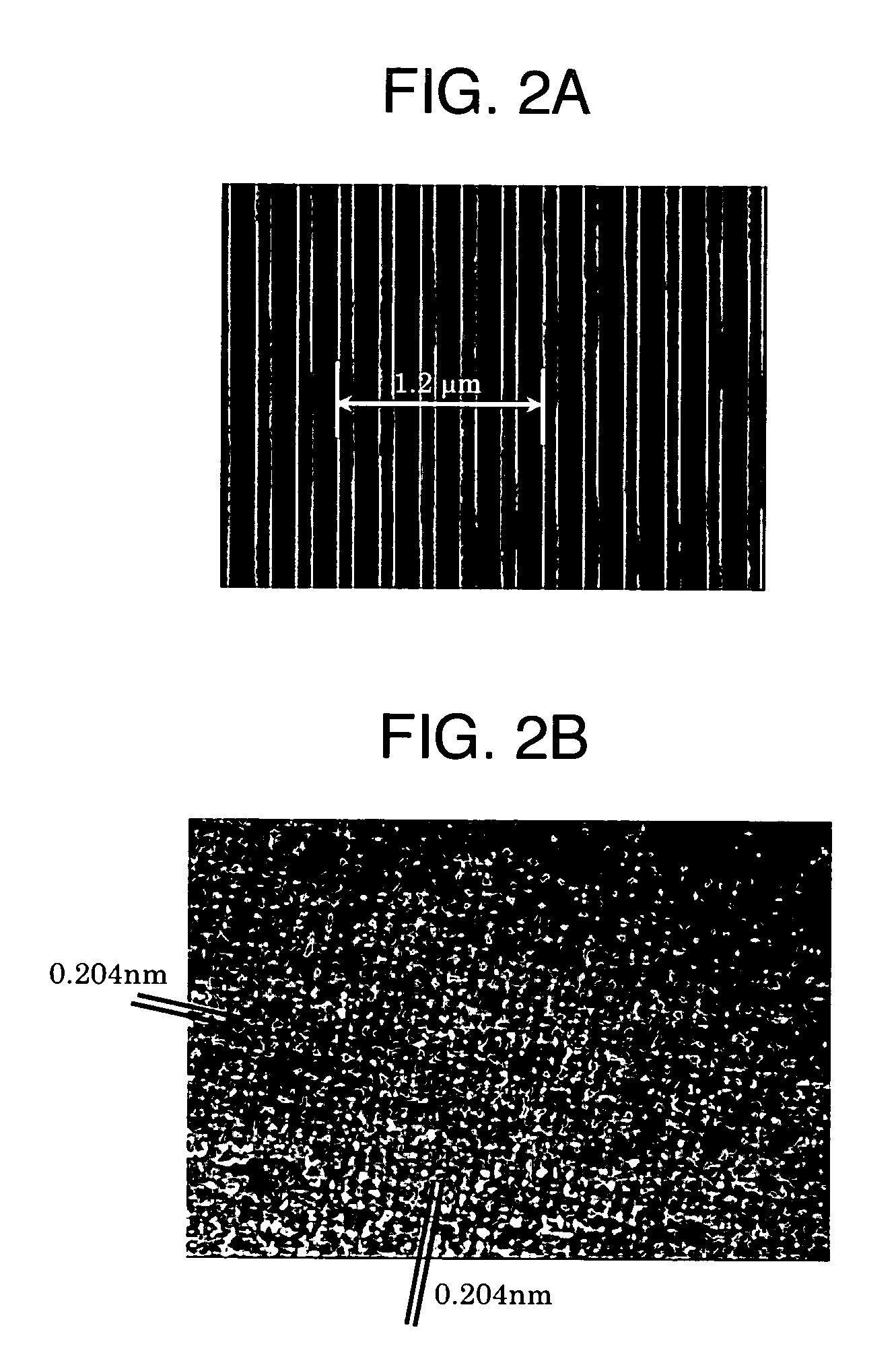
| Property | Measurement | Unit |
|---|---|---|
| pitch distance | aaaaa | aaaaa |
| crystal lattice spacing | aaaaa | aaaaa |
| lattice image | aaaaa | aaaaa |
Abstract
Description
Claims
Application Information