
Methods and apparatus for evaluating image focus

a technology of image focus and focusing method, which is applied in the field of focusing methods, can solve the problems of insufficient accuracy of focusing level, inability to achieve adequate focus, and inability to accurately identify abnormal tissue samples by direct visual observation alon

Inactive Publication Date: 2008-12-23
LUMA IMAGING CORP
View PDF364 Cites 60 Cited by
  • Summary
  • Abstract
  • Description
  • Claims
  • Application Information

AI Technical Summary

Benefits of technology

[0009]The invention provides methods of focusing an instrument for the acquisition of optical data from a tissue sample. Methods of the invention allow rapid focusing in the context of a diagnostic procedure in which rapid data acquisition is desirable. For example, inventive methods allow a user to focus an optical instrument quickly enough to obtain data within an optimal window of time following application of an agent to the tissue.
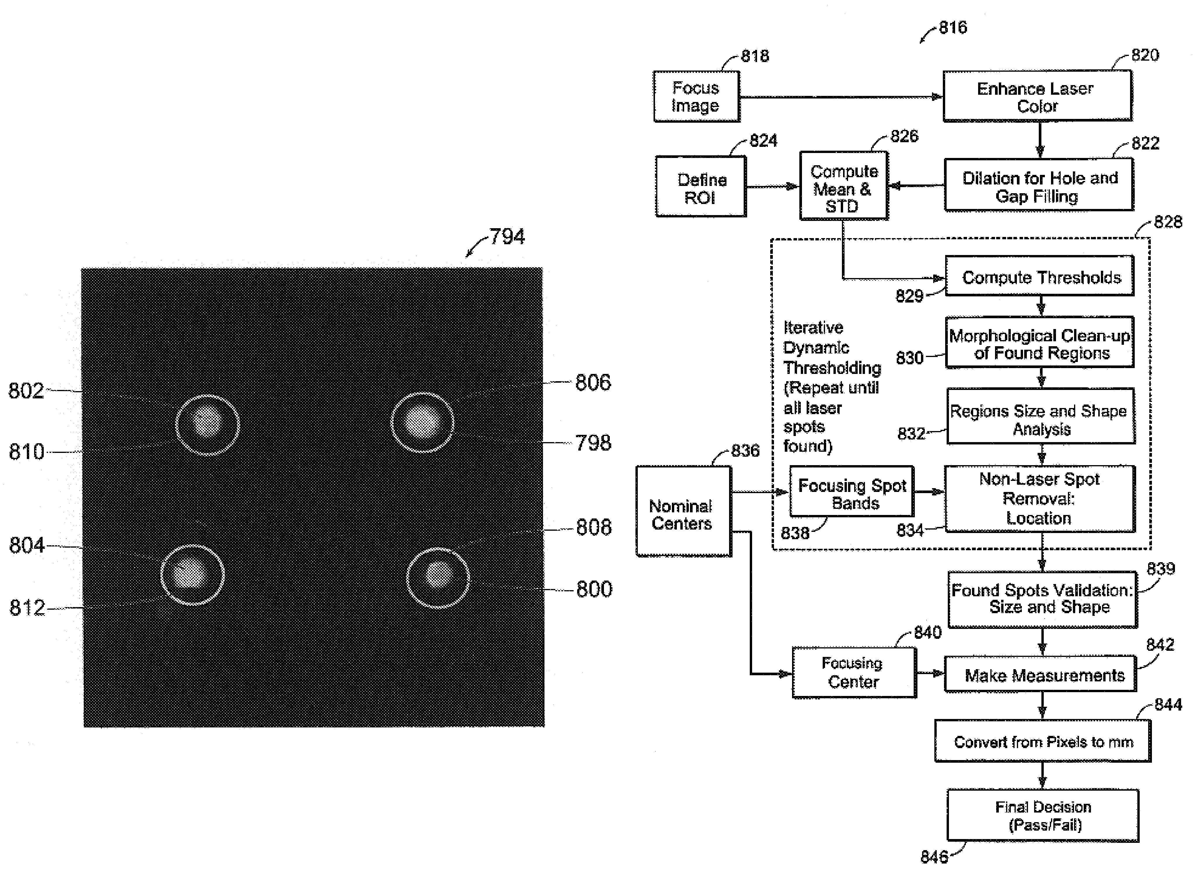
[0013]Obstructions on the surface of the tissue, as well as surface roughness and other irregularities, may distort the focusing spots projected onto the tissue. As a result, some of the focusing spots may not be clearly visible or may be distorted such that it is impossible to align all the spots within the superimposed focusing elements. Accordingly, preferred methods of the invention comprise automatic alignment validation to detect the locations of the light spots and to determine whether the spots are sufficiently well-aligned. Automatic validation offers an additional advantage in that it avoids delays caused by discovering improper focus after an optical scan has begun. Thus, automatic validation facilitates obtaining optical data within a prescribed, optimal window of time following application of contrast agent.

Problems solved by technology

However, direct visual observation alone may be inadequate for proper identification of an abnormal tissue sample, particularly in the early stages of disease.
However, current focusing methods generally do not provide sufficiently accurate levels of focus for acquiring diagnostic optical data from a tissue sample.
Tissue surface roughness, as well as obstructions such as glare, shadow, and blood, make achieving adequate focus difficult.
Even where adequate focus levels may be achieved, current focusing methods are generally not fast enough to allow acquisition of diagnostically-relevant optical data.
Furthermore, current focusing techniques are not sufficiently robust such that consistent focus levels are achieved over the lifetime of the optical instrument.

Method used


Image

  • Methods and apparatus for evaluating image focus
  • Methods and apparatus for evaluating image focus
  • Methods and apparatus for evaluating image focus

Examples


Embodiment Construction

[0164]

Table of ContentsPageSystem overview31Instrument36Spectral calibration50Patient scan procedure98Video calibration and focusing101Determining optimal data acquisition window113Motion tracking130Broadband reflectance arbitration and low-signal masking157Classification system overview179Spectral masking185Image masking196Glarevid202[ROI]vid206[ST]vid208Osvid215Bloodvid220Mucusvid224[SP]vid229[VW]vid240[FL]vid254Classifiers263Combining spectral and image data274Image enhancement283Diagnostic display289

[0165]The Table of Contents above is provided as a general organizational guide to the Description of the Illustrative Embodiment. Entries in the Table do not serve to limit support for any given element of the invention to a particular section of the Description.

[0166]System 100 Overview

[0167]The invention provides systems and methods for obtaining spectral data and image data from a tissue sample, for processing the data, and for using the data to diagnose the tissue sample. As use...


PUM

PropertyMeasurementUnit
timeaaaaaaaaaa
timeaaaaaaaaaa
optical dataaaaaaaaaaa

Abstract

The invention provides methods of focusing an instrument for the acquisition of optical data from a tissue sample. Methods of the invention allow rapid focusing in the context of a diagnostic procedure in which rapid data acquisition is desirable. For example, inventive methods allow a user to focus an optical instrument quickly enough to obtain data within an optimal window of time following application of a chemical agent to the tissue. In one embodiment, a user focuses an optical instrument by aligning laser spots projected onto a tissue sample within rings that are superimposed at predetermined locations within the user's visual field. Preferred methods of the invention further comprise automatic validation to detect whether the spots are sufficiently well-aligned.

Description

RELATED APPLICATIONS[0001]This application is related to the following commonly-owned applications: U.S. patent application Ser. No. 10 / 418,922, entitled, “Methods and Apparatus for Characterization of Tissue Samples”; U.S. Pat. No. 7,136,518, entitled, “Methods and Apparatus for Displaying Diagnostic Data”; U.S. patent application Ser. No. 10 / 418,974, entitled, “Methods and Apparatus for Visually Enhancing Images”; U.S. Pat. No. 7,309,867, entitled, “Methods and Apparatus for Characterization of Tissue Samples”; U.S. patent application Ser. No. 10 / 418,975, entitled, “Methods and Apparatus for Processing Image Data for Use in Tissue Characterization”; U.S. Pat. No. 7,282,723, entitled, “Methods and Apparatus for Processing Spectral Data for Use in Tissue Characterization”; and U.S. patent application Ser. No. 10 / 418,973, entitled, “Methods and Apparatus for Calibrating Spectral Data,” all of which are filed on even date herewith.FIELD OF THE INVENTION[0002]This invention relates gen...

Claims


Application Information

Patent Timeline
Patent Type & Authority Patents(United States)
IPC IPC(8): A61B6/00A61B5/00G01N21/31
CPCA61B5/0059G01N21/31A61B5/7264A61B5/7257A61B5/7267G16H50/20
Inventor BANKS, PERRY S.JIANG, CHUNSHENGTWIETMEYER, KAREN M.MORRELL, RICHARD M.ABELE, CHARLES C.
Owner LUMA IMAGING CORP
Learn More