Method of preparing a packaged antimicrobial medical device
a medical device and packaging technology, applied in the field of packaging antimicrobial medical devices, can solve the problems of significant increase in the cost of treatment for patients, infection and trauma to patients, etc., and achieve the effects of inhibiting bacterial colonization, inhibiting bacterial growth, and inhibiting bacterial colonization
- Summary
- Abstract
- Description
- Claims
- Application Information
AI Technical Summary
Benefits of technology
Problems solved by technology
Method used
Image
Examples
example 1
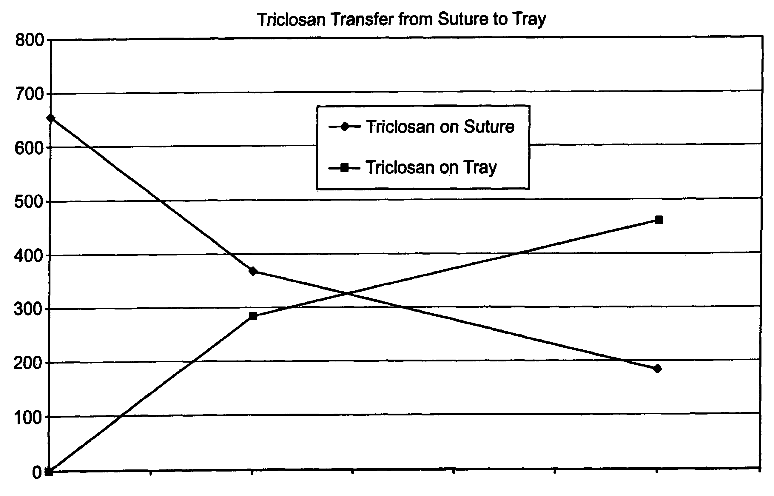
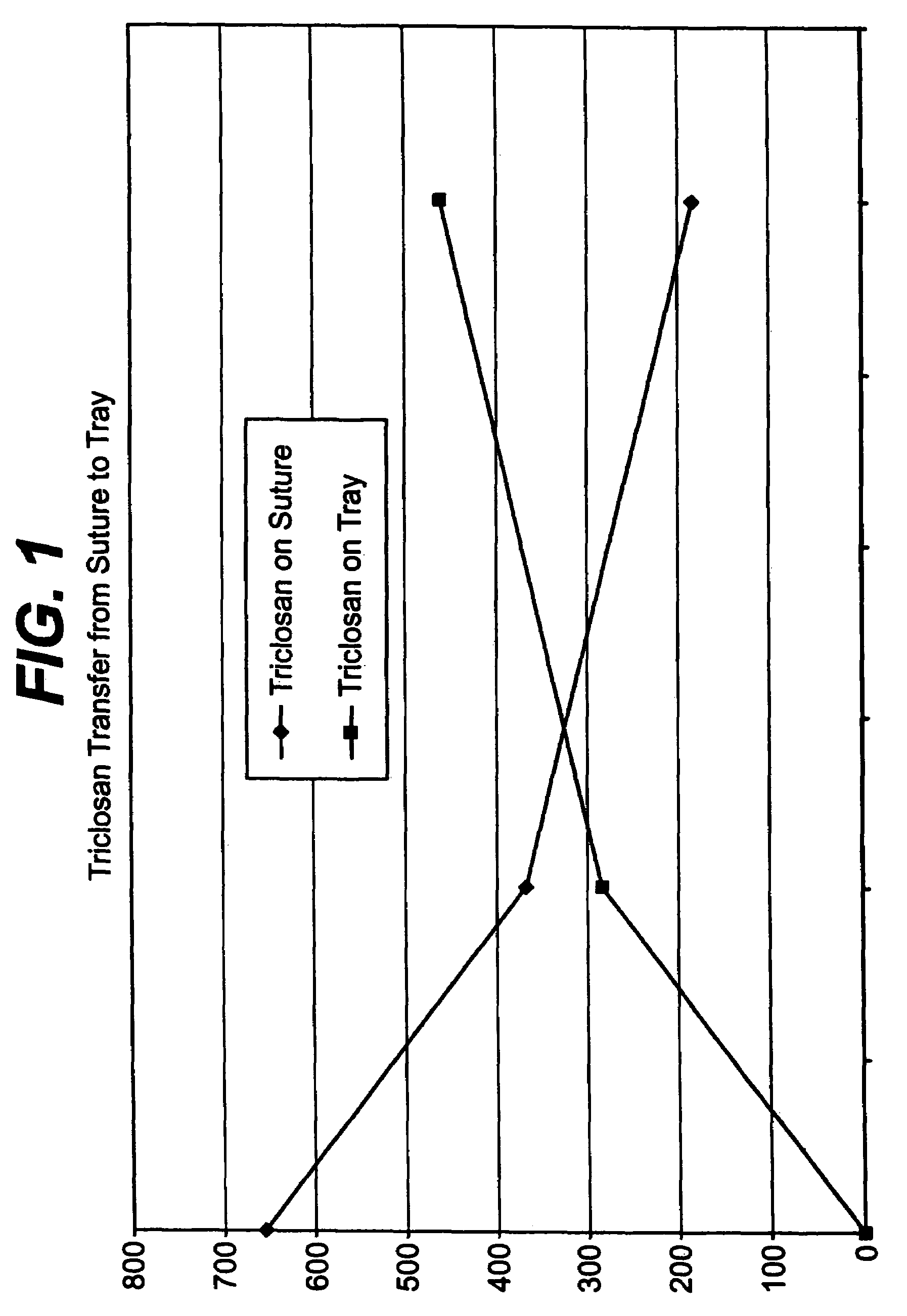
[0043]A series of USP standard size 5-0 coated polyglactin 910 sutures were coated with a 2% triclosan coating composition so that each suture contained about a total of 23.2 μg triclosan before sterilization. The coated sutures each were placed in a package as described herein above including a containment component, i.e., a tray, for holding the suture and a paper component for covering the suture in the tray. The suture in the containment component and packaging were sterilized as described herein above. After sterilization, it was determined that that suture contained about 5.5 μg triclosan, the tray about 0.2 μg triclosan, the paper component about 2.3 μg triclosan, and the package heat seal coating about 1.5 μg triclosan. Triclosan not recovered after sterilization was about 13.7 μg triclosan. FIG. 1 indicates triclosan transfer from the antimicrobial suture to the tray of the package as a function of time at 55° C.
[0044]After sterilization, the paper component and tray of the...
example 2
[0048]This example is a 24-hour aqueous immersion assay. The purpose of this assay was to determine the effect of aqueous exposure on the antimicrobial properties of suture material for a range of suture diameters. Sterile sutures in USP sizes 2-0, 3-0, 4-0, and 5-0, with and without a 1% triclosan coating applied thereto, were aseptically cut into 5-cm pieces. One half of the cut pieces were stored in a sterile Petri dish and kept under a dry nitrogen atmosphere for 24 hours (dry suture). One half of the cut pieces were aseptically transferred to sterile 0.85% saline and incubated at 37° C. for 24 hours (wet sutures).
[0049]The dry and wet sutures were then aseptically placed in individual sterile Petri dishes and challenged with 100 microliters of inoculum containing 105 colony-forming units (CFU) of Staphylococcus aureus or Staphylococcus epidermidis. Ten replicates of each suture size were used for each organism and for both the dry and wet sample groups. TSA was poured into each...
example 4
[0053]This example is directed to a 7-day aqueous immersion assay. The purpose of this assay was to determine if the antimicrobial effect of triclosan treatment would endure for 7 days in a buffered aqueous environment.
[0054]Sterile USP size 2-0 coated polyglactin 910 suture coated with a 1%, 2%, and 3% triclosan coating solution, respectively, and ethylene oxide sterilized USP size 2-0 coated polyglactin suture were aseptically cut into 5-cm pieces. Samples were tested on each of 7 days in triplicate.
[0055]On day 1, 3 pieces of each suture material were placed into individual sterile Petri dishes and inoculated with 0.1 mL of challenge organism containing approximately 104 CFU. TSA was poured into each dish and allowed to solidify. All remaining pieces of suture material were placed into 100 mL of sterile phosphate buffered 0.85% saline (PBS). Every 24 hours for the next 6 days, 3 pieces of each suture material were removed from the PBS, inoculated, and pour plated in tryptic / soy / a...
PUM
| Property | Measurement | Unit |
|---|---|---|
| pressure | aaaaa | aaaaa |
| temperature | aaaaa | aaaaa |
| temperature | aaaaa | aaaaa |
Abstract
Description
Claims
Application Information
 - R&D
- Intellectual Property
- Life Sciences
- Materials
- Tech Scout
- Unparalleled Data Quality
- Higher Quality Content
- 60% Fewer Hallucinations
Browse by: Latest US Patents, China's latest patents, Technical Efficacy Thesaurus, Application Domain, Technology Topic, Popular Technical Reports.
© 2025 PatSnap. All rights reserved.Legal|Privacy policy|Modern Slavery Act Transparency Statement|Sitemap|About US| Contact US: help@patsnap.com


